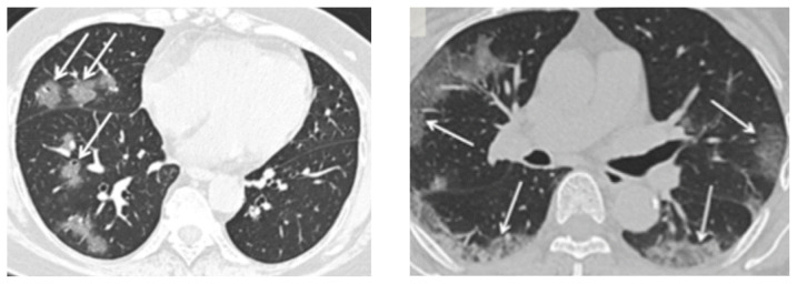
Figure 7.
CT Scans with Typical Findings in COVID-19 Patients
Left: An axial CT image shows ground-glass opacities with a rounded morphology in the right middle and lower lobes.
Right: An axial CT image shows bilateral ground-glass and consolidative opacities with a peripheral distribution.
Credit: Department of Radiology, Icahn School of Medicine at Mount Sinai, New York. Reproduced with permission.

