FIG 1.
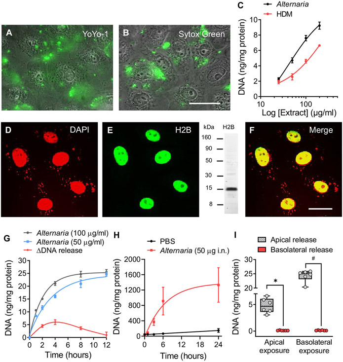
Alternaria stimulates DNA release. Fluorescence/phase contrast images (original magnification 40×, scale bar = 10 μm, n = 3) showing eDNA with (A) YoYo-1 (1 μmol) and (B) SYTOX Green (10 μmol) after Alternaria (30 minutes). (C) Concentration effects of Alternaria and HDM on eDNA (Alternaria, half maximal effective concentration [EC50] = 42 μg/mL; r2 = 0.89 and HDM, EC50 = 274 μg/mL; r2 = 0.90). (D) DAPI-labeled nuclei 30 minutes after Alternaria (100 μg/mL) treatment (n = 3). (E) Histone 2B (H2B) localization 30 minutes after Alternaria (100 μg/mL) exposure. Western blot showing H2B labeling with antibodies used for immunocytochemistry. (F) Colocalization (yellow) of DNA and H2B in the nucleus (original magnification 60×, scale bar = 5 μm). (G) DNA release at 2 Alternaria concentrations (50 and 100 μg/mL), n = 6 each. ΔDNA release (red) represents differences in DNA accumulation between time intervals after Alternaria (100 μg/mL) exposure. (H) eDNA in BAL fluid of mice (n = 5) treated intranasally with Alternaria (50 μg). (I) Apical and basolateral treatment of 16HBE14o− monolayers exposed to 100 μg/mL Alternaria for 30 minutes (n = 6 each, #P < .0004, *P < .0001; unpaired, 2-tailed ttests with Welch correction).

