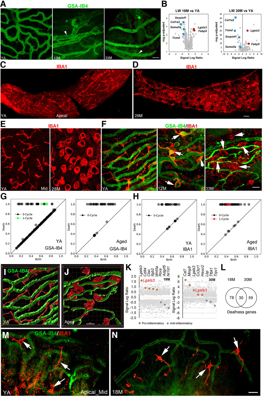
Figure 2.
Age-related strial microvasculature and macrophage alterations and associated gene expression changes in middle-aged and aged CBA/CaJ mice. A, Isolectin GSA-IB4 (from Griffonia simplicifolia, Alexa Flour 488 conjugate) stained strial capillaries in the apical turn of young adult (YA; left panel), middle-aged (middle panel), and aged (right panel) mice. Enlarged capillaries (arrowheads) were seen in the apical SV in both middle-aged and aged mice. B, Volcano plots of vascular function genes in the LW of middle-aged (left panel) and aged (right panel) mice (Extended Data Fig. 2-1). Selected upregulated (red) and downregulated (blue) genes are highlighted; values for other genes are shown in gray. Note that Lgals3 was identified as differentially expressed in the LW of both middle-aged and aged CBA/CaJ mice. C–E, Macrophages in the SV undergo dramatic alterations in morphology in aged mice. F, High-resolution images reveal age-related changes in macrophage morphology and macrophage-capillary interactions in the SV of middle-aged (middle panel) and aged mice (right panel). G, H, Persistent homology analyses were performed using images of whole-mount preparations (right and left panels in F) to quantify the distinct GSA-IB4+ microvascular (G) and IBA1+ macrophage (H) structural profiles in YA and aged mice. Diagrams show more one-cycle loop structures in the microvasculature of YA compared with aged mouse. In contrast, the increase in one-cycle loops in the aged mouse (H, left panel) indicates a greater number of activated macrophages with ringed ameboid appearance compared with YA (H, right panel). The x- and y-axes show relative units after scaling to maximal death value from each analysis. I, J, 3D reconstruction images generated by Imaris 3D volume rendering highlight morphologic changes in macrophages (red) of aged mice that are associated with strial microvasculature (green). Images were generated from Z-stacks of F, left and right panels, respectively. K, Transcriptomic data for LW from middle-aged and aged mice as compared with YA controls. Genes related to macrophage activation that are proinflammatory (red) or anti-inflammatory (blue) were differentially expressed in middle-aged (left panel) and aged (right panel) LW (see detailed information in Extended Data Fig. 2-2). L, Genes linked to human deafness are differentially expressed in middle-aged and aged LW (see detailed information in Extended Data Fig. 2-3). The reference list of ∼700 genes involved in human deafness was obtained from Lewis et al. (2022). M, N, IBA1+ macrophages (arrows) in the AN region show no significant changes in the middle-aged cochleas compared with cochleas in young adult mice. Scale bars: 20 µm in A; 30 µm in C; 20 µm in F; and 20 µm in J and N (applies to I, J, M, N).

