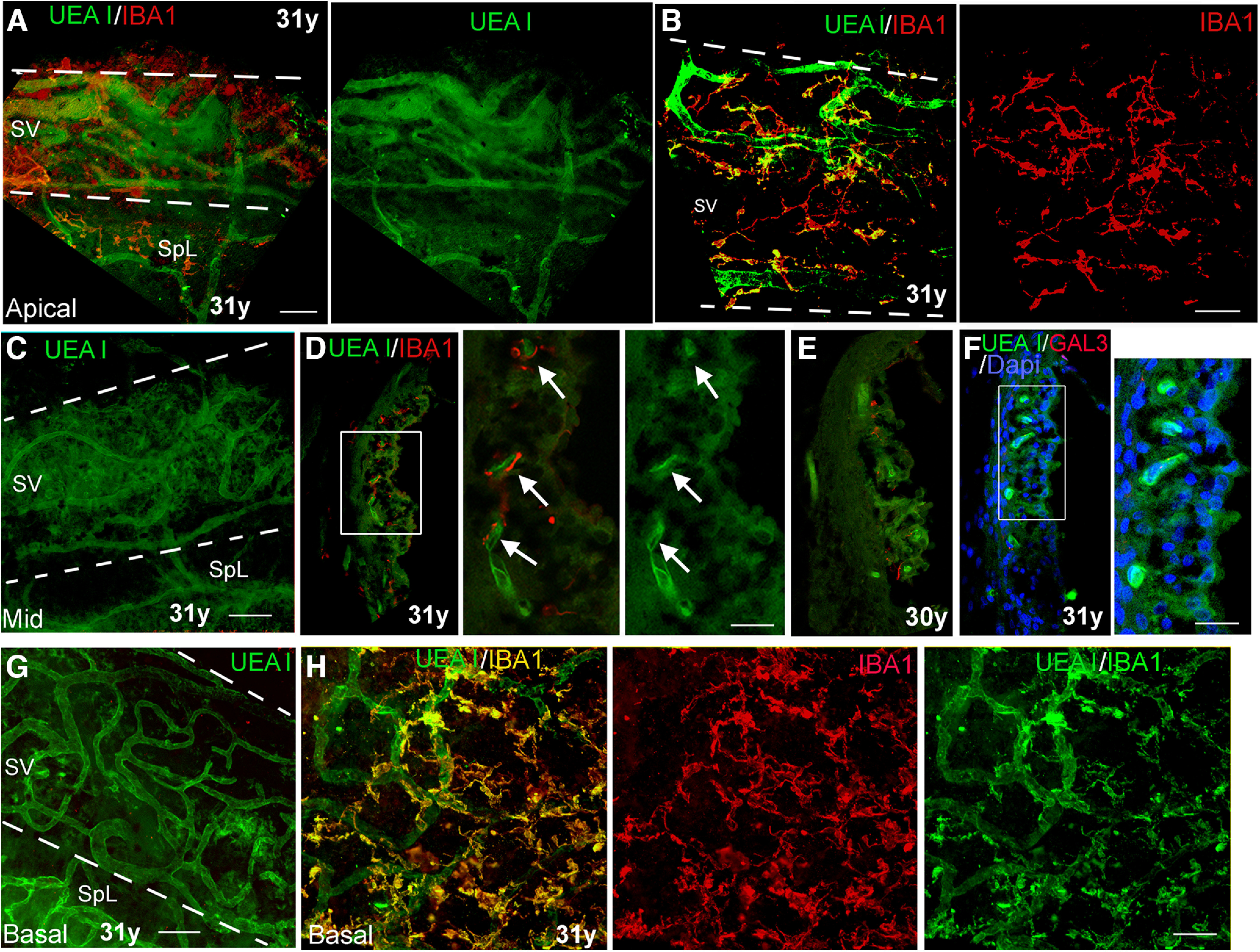
Figure 5.

A lectin histochemical approach to visualize interactions between macrophages and SV microvasculature in cochleas from human temporal bones from young adult donors. A, B, Dual staining with UEA I and anti-IBA1 in a whole-mount preparation of a temporal bone from a young adult donor. The images taken from the apical turns (A, B) revealed IBA1+ macrophages (red) and UEA I+ microvasculature network. The strial area is highlighted with two dotted lines. SpL, spiral ligament. C, UEA I+ microvasculature network in the middle turn of the temporal bone from the same donor. D, E, Dual staining of UEA I and anti-IBA1 on the sections of temporal bones from young adult donors clearly show the cellular processes of the IBAI+ macrophages closely opposed to UEA I+ strial microvasculature (arrows). D, Central panel, Enlarged image of the boxed area in the left panel. F, No strial cells stained positively with the macrophage activation marker GAL3. F, Right panel, Enlarged image of the boxed area in F. Note that there are GAL3+ cells in the SV from the temporal bones from middle-aged and older donors (see Fig. 6). G, UEA l+ microvasculature in the SV of the basal turn from the temporal bone from a young adult donor. H, Interactions of IBA1+ macrophages (yellow) with UEA l+ microvasculature (green) were visualized in the basal turn from the temporal bone from a young adult donor. Scale bars: 50 µm in A–C, G, H; 20 µm in right panels in D, F.
