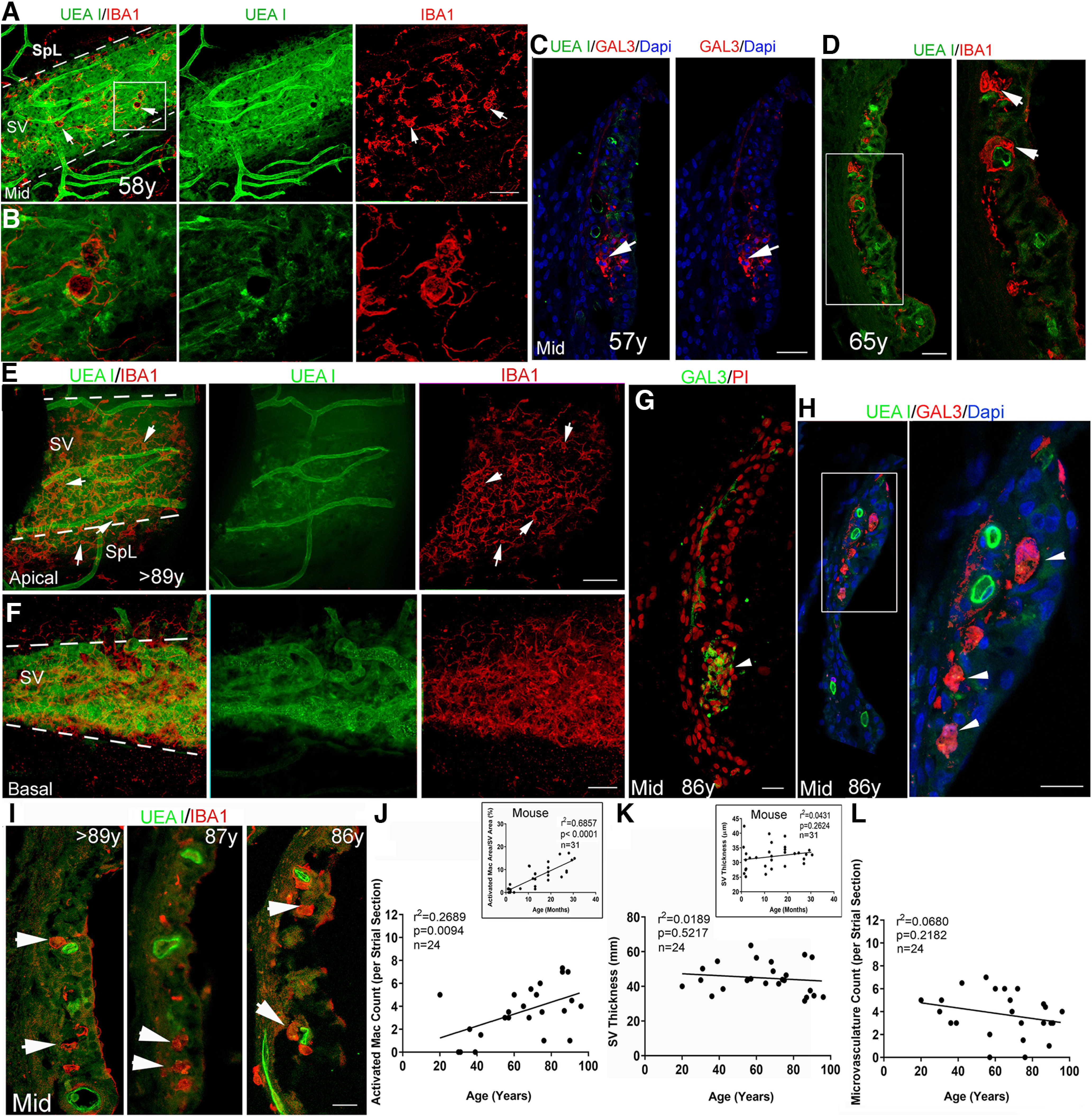
Figure 6.

Increased macrophage activation in the SV from temporal bones from middle-aged and older donors. A, B, Dual staining with UEA l and anti-IBA1 in whole-mount preparations in a temporal bone from a middle-aged donor. The image from the middle turn reveals increased IBA1+ macrophage (red) activation and a disrupted interaction of macrophages with the UEA l+ microvasculature (green). Strial area is highlighted by two dotted lines. B, Enlarged image of the boxed area in A. C, GAL3+ cells (arrows) in the SV of the LW in a temporal bone from another middle-aged donor. D, IBA1+ macrophages with an amoeboid shape (arrows) were seen in the SV of a temporal bone from a middle-aged donor. Right panel, Enlarged image of the boxed area in the left panel. E, F, Dual staining with UEA l and anti-IBA1 in whole-mount preparations from a temporal bone from an older donor. Images from the LW in the apical (E) and basal (F) turn revealed a reduction of UEA l+ microvasculature in the apical turn. This age-related strial microvasculature difference was confirmed using the persistent homology approach (Fig. 7). Increased IBA1+ macrophage (red) activation was seen in both apical and basal turns. Arrows identify macrophages with an amoeboid-shaped cell body and fewer elongated cellular processes. G, H, GAL3+ cells (arrows) were identified in the SV of the LW section in the temporal bone from another older donor. I–L, An increase in IBA1+ macrophages with an amoeboid shape (arrowheads) is seen in the SV from temporal bones from three older donors compared with that in temporal bones from young adult donors in Figure 5D,E. This observation is supported by linear regression analyses, which revealed a significant age-dependent increase in activated macrophages in the middle turn SV (main image in J). There was no significant correlation between UEA I+ microvasculature count in the middle turn and age at donation (p > 0.05). Scale bars: 50 µm in A; 50 µm in E, F; 20 µm in C, D, G, H, right panel, I.
