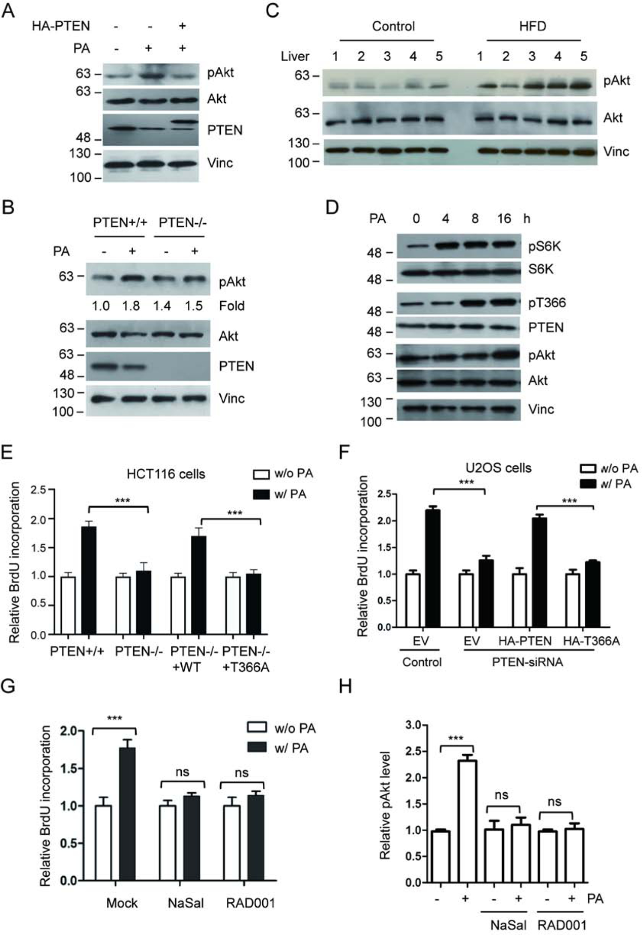
Figure 4.

PA treatment promotes cancer cell proliferation. (A) U2OS cells were treated with PA, with or without PTEN overexpression, and activation of Akt was detected using anti-Akt p-S473 antibody (pAkt). (B) HCT116 PTEN+/+ or PTEN−/− cells were exposed to PA and Akt, pAkt and PTEN were detected with corresponding antibodies. (C) Activation of Akt in liver samples for five HFD-fed mice and five control mice was determined with anti-Akt pS473 antibody (pAkt). (D) U2OS cells were treated with 400 mM PA. Following the treatment the phosphorylation levels of S6K, PTEN, or Akt were detected by IB with corresponding antibodies at the indicated times. (E) HCT116 PTEN+/+ or PTEN−/− cells were treated with control or 400 mM PA, transfected with wild type PTEN or T366A, and cell proliferation was determined by measuring the incorporation of BrdU into DNA. (F) U2OS cells, transfected with PTEN-siRNA, wild type PTEN or T366A as indicated, were treated with control or 400 mM PA. Cell proliferation was determined by measuring the incorporation of BrdU into DNA. (G, H) U2OS cells were treated with control or 400 mM PA in the presence or absence of RAD001 or NaSal. Cell proliferation was measured by incorporation of BrdU (G) and Akt phosphorylation levels were determined by IB and normalized to Akt protein levels (H). ***P<0.001, One-way ANOVA.
