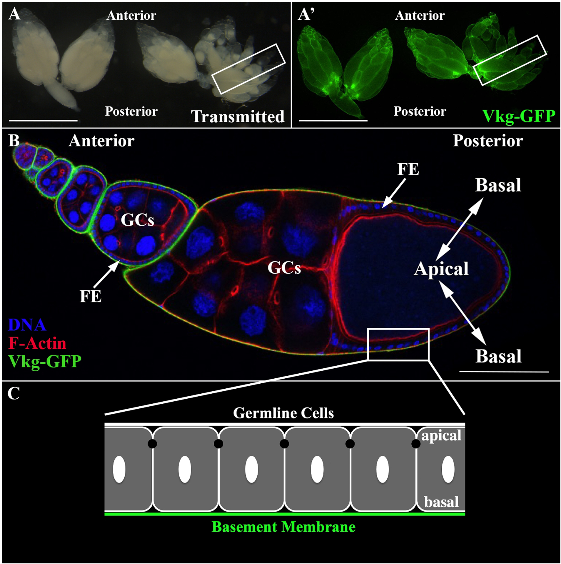
Figure 1: The follicular epithelium (FE) of the Drosophila ovary: a model system to study the polarized deposition of basement membrane (BM) proteins.

(A) Image of intact ovaries after dissection and excision taken with a fluorescence stereomicroscope. Ovaries are expressing an endogenous GFP-tagged BM protein (Vkg-GFP). Scale bar = 1 mm. (A’) Two ovaries of a female fly are attached at the oviduct. Each ovary contains 16–20 ovarioles. A single ovariole is outlined (rectangle). Scale bar = 1 mm. (B) Longitudinal section, taken with a confocal microscope, through an ovariole expressing Vkg-GFP and stained for DNA (blue) and F-Actin (red). Ovarioles consist of egg chambers at different stages. Egg chambers are composed of a monolayer follicular epithelium (FE) that surrounds the germline cells (GCs). The FE synthesizes and basally secretes BM proteins (e.g., Pcan and Vkg). Scale bar = 100 μm. (C) Schematic of the FE. The FE is a classic epithelium with a distinct apical-basal polarity where the apical domain faces the germline cells, and the basal domain faces the BM (green).
