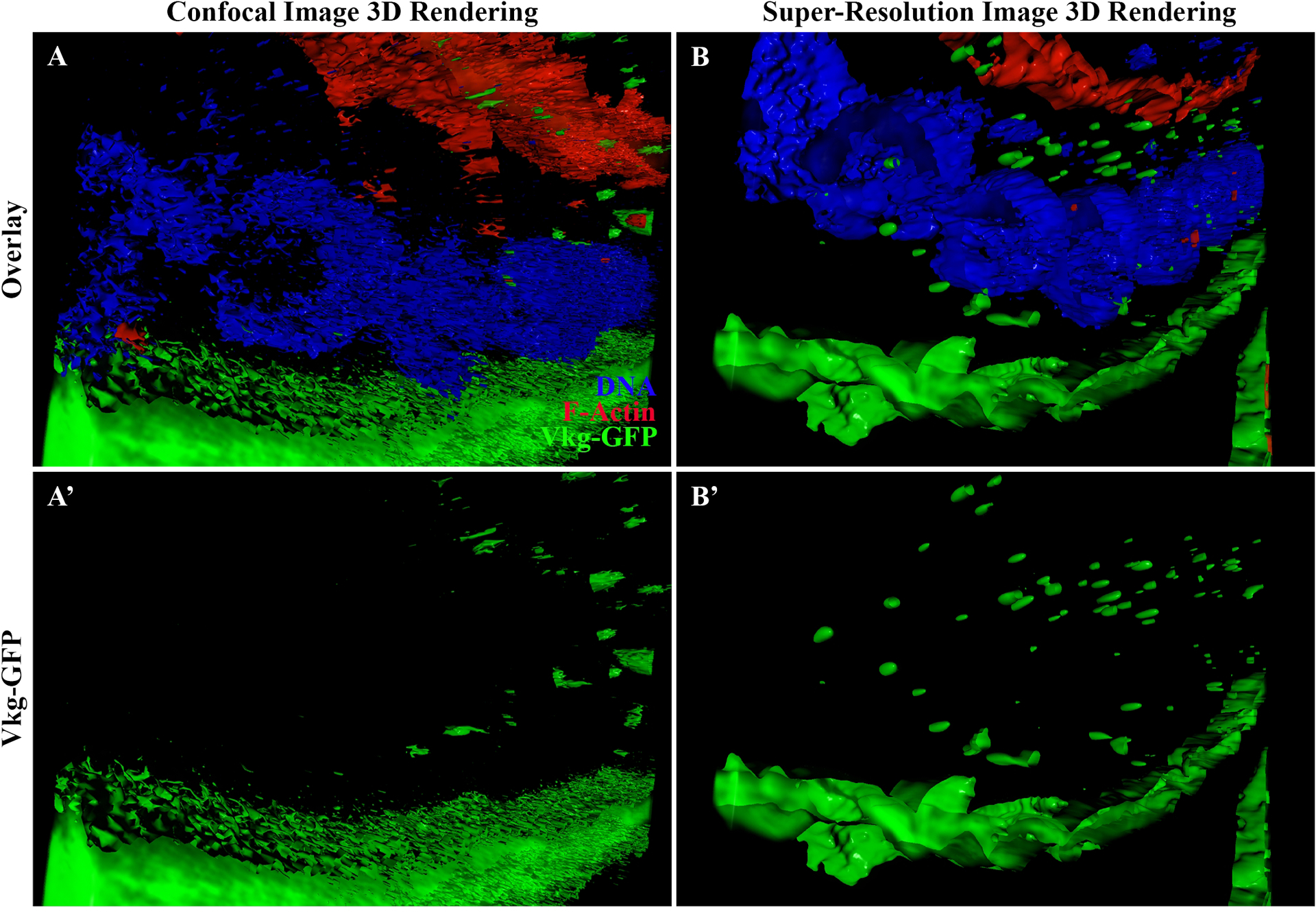
Figure 5: 3D reconstruction of a z-stack acquired and processed using confocal and super-resolution microscopy approaches.

(A-B) 3D rendering of z-stacks (mixed view) of egg chambers expressing Vkg-GFP (green) and stained for DNA (blue) and F-actin (red). (A) 3D rendering of a z-stack acquired via confocal microscopy. The location and shape of compartments and vesicles containing Vkg-GFP can be seen throughout the cells (A’). (B) 3D rendering of a z-stack acquired via optimal super-resolution processing. The location and shape of compartments and vesicles containing Vkg-GFP can be seen (B’). The shape of vesicles, as well as the BM and the nuclei, are smoothly defined, and the resolution is higher compared to that of confocal microscopy (compared B’ and A’). The shape and size of the compartments and vesicles containing Vkg-GFP can also be better determined using super-resolution microscopy (compared B’ and A’).
