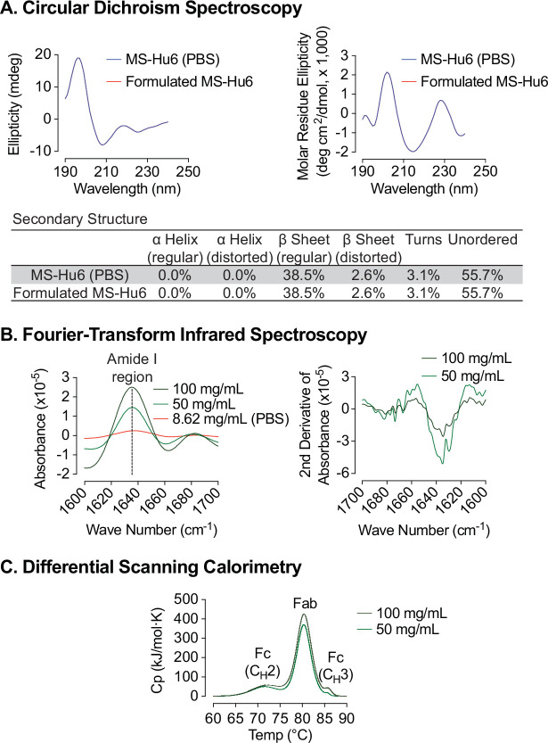
Figure 4. Biophysical characterization of formulated MS-Hu6.
Circular dichroism (CD) spectroscopy evaluated the secondary structure of formulated MS-Hu6. Analysis in the far UV region (190–240 nm) revealed that the predominant secondary structure in MS-Hu6 was regular β-sheets and unordered/random coils (A). Secondary structure was also confirmed at higher formulation concentrations (50 and 100 mg/mL) using Fourier–transform infrared (FTIR) spectroscopy. The amide I band peak at 1637 cm–1 (intra–molecular β-sheets) and the random coil (1642–1657 cm–1) did not shift, confirming maintenance of the native conformation in formulation (B). Thermostability was further confirmed using nano differential scanning calorimetry (Nano DSC). MS-Hu6 concentrations of 50 and 100 mg/ml had comparable Tms, indicating that the overall structure is conformationally and thermally stable at high concentrations (C).

