. 2023 Jun 22;12:e85432. doi: 10.7554/eLife.85432

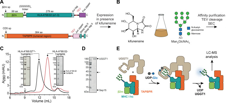
Figure 1. In-vitro system of UGGT1-catalyzed quality control and reglucosylation of MHC I.

(A) Construct design to obtain peptide-receptive HLA-A*68:02-TAPBPR complexes by transient co-transfection of HEK293-F cells. (B) Co-expression of HLA-A*68:02 and TAPBPR was performed in the presence of the α-mannosidase I inhibitor kifunensine to generate a defined Man9GlcNAc2 glycan tree on the MHC I that can be recognized by UGGT1. (C) The secreted leucine zippered complex was isolated from the cell culture supernatant by Strep-Tactin affinity purification. Leucine zippers and the Twin-Strep-tag of TAPBPR were removed by protease treatment. The MHC I-chaperone complexes were further analyzed and purified by size exclusion chromatography (SEC) on a Superdex 200 (Increase 10/300) column. (D) SDS-PAGE analysis of immobilized metal-affinity chromatography (IMAC)-purified human wildtype (wt) UGGT1 co-expressed with Sep15 and secreted from insect cells. The asterisk (*) indicates a degradation product of UGGT1. (E) The purified UGGT1-Sep15 complex was employed in the reglucosylation assay with the peptide-receptive HLA-A*68:02-TAPBPR complex of (C) harboring the high-mannose glycan on MHC I. Abbreviations: A280: absorption at 280 nm; aa: amino acids; kDa: kilodalton; mAU: milli-absorption units; MHC I hc: MHC I heavy chain; sp: signal peptide; UDP-Glc: UDP-glucose; V0: void volume.

Figure 1—source data 1. Original SDS-PAGE gel of HLA-A*68:02Fos-TAPBPRJun, Figure 1C.
Figure 1—source data 2. Original SDS-PAGE gel of HLA-A*68:02-TAPBPR, Figure 1C.
Figure 1—source data 3. Original SDS-PAGE gel of UGGT1-Sep15, Figure 1D.

Figure 1.

Figure 1—figure supplement 1. Influence of kifunensine concentration on the proportion of Man9GlcNAc2 among the Man7-9GlcNAc2 MHC I glycan species when co-expressed with TAPBPR in HEK293-F cells.
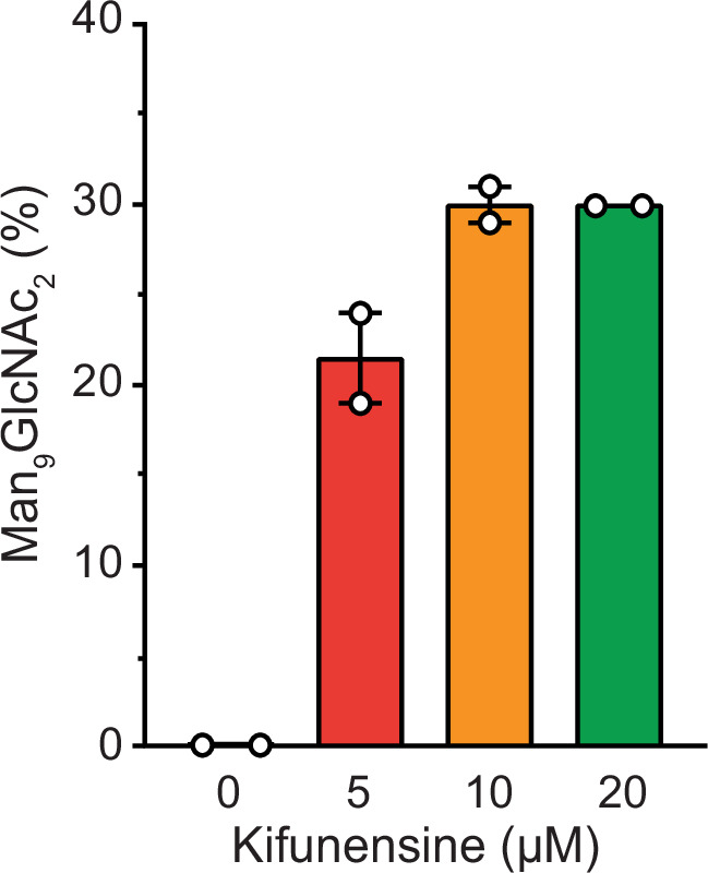
Figure 1—figure supplement 1.

The glycan status of TAPBPR-bound HLA-A*68:02 was analyzed by LC-MS after expression in presence of various concentrations of kifunensine. Proportion of Man9GlcNAc2 was calculated based on the total glycan intensity (Man7-9GlcNAc2). Data represent mean ± SEM (n=2) from two independent protein expressions and purifications.
Figure 1—figure supplement 2. Analysis of purified human UGGT1.

Figure 1—figure supplement 2.

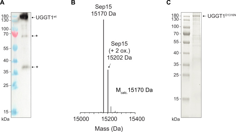
(A) Anti-UGGT1 immunoblot analysis after immobilized metal-affinity chromatography (IMAC) purification of wildtype (wt) human UGGT1 co-expressed with Sep15 and secreted from insect cells. The asterisks (*) indicate degradation products of UGGT1. (B) Compositional analysis of purified UGGT1wt-Sep15 by liquid chromatography-mass spectrometry (LC-MS) (deconvoluted spectrum). The voltages and desolvation temperature were adjusted for optimal ionization of the 15 kDa protein Sep15, precluding simultaneous efficient ionization of the 175 kDa protein UGGT1 within the same measurement. (C) Non-reducing SDS-PAGE analysis of the catalytically inactive UGGT1D1316N mutant expressed in HEK293-F cells and purified by IMAC.
Figure 1—figure supplement 2—source data 1. Original anti-UGGT1 immunoblot, Figure 1—figure supplement 2A.
Figure 1—figure supplement 2—source data 2. Original SDS-PAGE gel of UGGT1D1316N, Figure 1—figure supplement 2C.