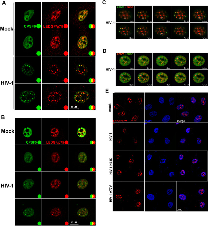
Figure 8.
HIV-1 infection induces nuclear redistribution of LEDGF/p75. A549 cells were infected or not (mock) with wild-type HIV-1-Luc (A–D) or with HIV-1-GFP viruses bearing the capsid mutations N74D and A77V (E) at an MOI of ~ 2 for 24 h. Cells were fixed, permeabilized, and stained using rabbit polyclonal antibody to LEDGF/p75 (E) with mouse monoclonal antibody to CPSF6 (A and C) or with mouse monoclonal antibody to CPSF5 (B and D). Secondary antibodies were Alexa-488-conjugated donkey anti-mouse IgG (green channel) and Alexa-594-conjugated donkey anti-rabbit IgG (red channel). Nuclei were stained with DAPI (channel blue). Stained cells were examined by fluorescence microscopy. Merging the red and green channels generated the yellow color that indicates overlapping localization of these channels. Merging the red and blue channels generated the magenta color that indicates overlapping localization of these channels. For C and D z-stack images at 0.2 μm intervals were acquired. The white circle indicates an example of a condensate that contains CPSF6 (C) or CPSF5 (D) surrounded by LEDGF/p75. Scale bar = 10 μm.

