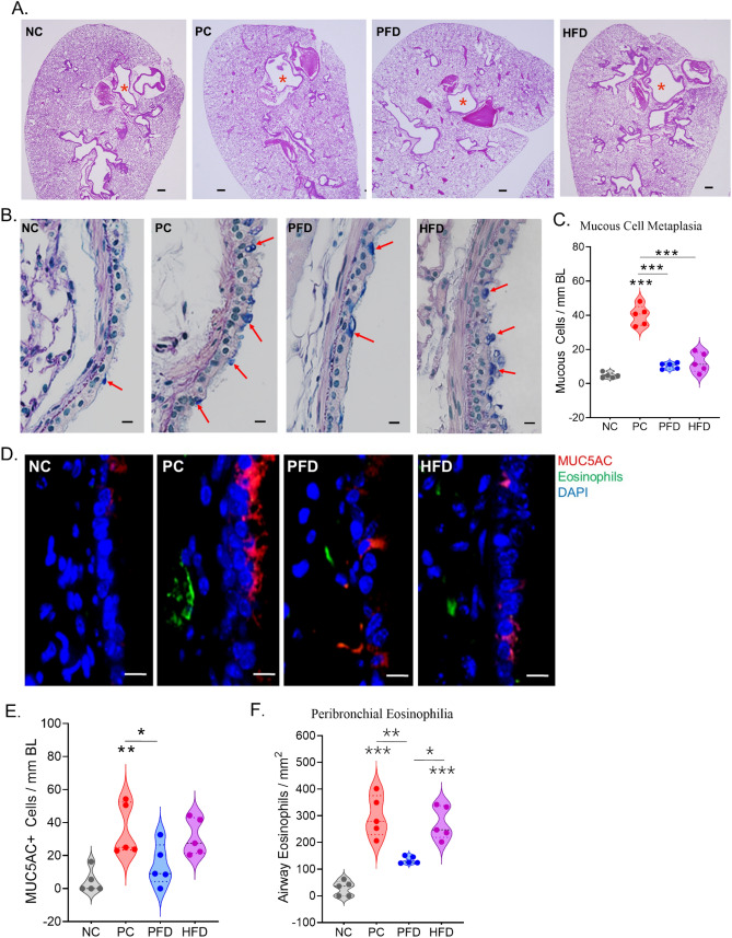
Figure 4.
The airway mucous cell metaplasia and peribronchial eosinophilia caused by CDE aerosol exposure is mitigated by PFD. Representative micrographs of mouse lung tissue from each group showing gross morphology and airway mucous metaplasia. (A) Low magnification images of whole lung section stained with H&E showing the bronchial airways (marked with a red asterisk), scale—200 µm. (B) A high magnification image of lung axial airways stained with AB/PAS showing the AB + mucin glycoprotein marking the mucous/goblet cells (marked with red arrows), scale—10 µm. (C) The Quantitation of mucous cells per mm of Basal lamina (BL). (D) Representative micrographs showing airway Muc5ac + mucous/goblet cells (shown in red) and the eosinophils (shown in green) with nuclei stained with DAPI (shown in blue), scale bar—10 µm. Quantitation of (E) Muc5ac + cells per mm of BL and (F) Eosinophils per mm2 area in each group of mice. Data shown as mean ± SEM and analyzed by ANOVA (n = 5/gp). *p < 0.05; **p < 0.01; ***p < 0.001.

