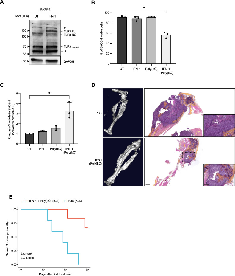
Fig. 2. TLR3 can mediate sarcoma cell death in vitro and in vivo.
A Treatment with IFN-1 is sufficient to restore TLR3 expression in SaOS-2 osteosarcoma (OS) cells. Immunoblot against TLR3 was performed on cell lysates, 24 h after IFN-1 treatment. GAPDH is used as a loading control. One representative image out of 3 independent experiments is shown. FL Full Length, NG Non-Glycosylated. Asterisks denote unspecific bands. B Activation of TLR3 by Poly(I:C) triggers SaOS-2 cell death in vitro. Viability of SaOS-2 osteosarcoma cells treated or not with IFN-1 and/or Poly(I:C) for 72 h was analyzed by cytometry (DAPI/Acridine Orange). Mean values ± s.t.d are represented (n = 3, *p-value = 0.05). C Activation of TLR3 by Poly(I:C) leads to an increase in Caspase-3 activity in SaOS-2 cells in vitro. Mean values ± s.t.d of caspase-3 activity quantified according to the fluorescence emitted by cleavage of a DEVD-AFC peptide are represented (n = 3, *p-value = 0.05). D, E Activation of TLR3 slows osteosarcoma progression in vivo. D Left panel. CT-Scan imaging of mice tibia/fibula bones, 15 days after SaOS-2 osteosarcoma cell engraftment. One representative image is shown for each group. Right panel. Representative images of Hematoxylin Phloxin and Safran staining showing dense, poorly differentiated, high-grade cells in a PBS treated mice (n = 5), and a smaller tumor in an IFN-1+Poly(I:C) treated animal (n = 6). Black scales: 1 mm (low magnification)/400 μm (high magnification). Mouse overall survival is represented as (E) a Kaplan–Meier curve (p-value = 0.0036, log-rank test).

