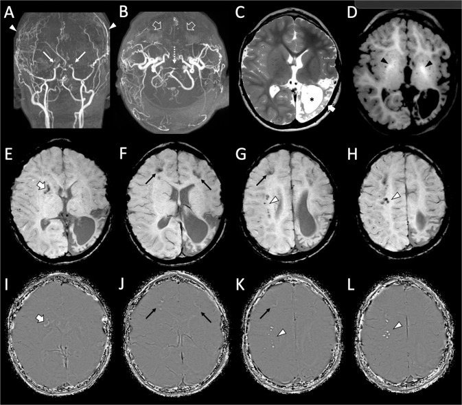
Fig. 1. Brain MR angiography and MRI of the patient performed at 4 years and 3 months of age.
A, B Arterial MR angiography images reveal very severe moyamoya vasculopathy with bilateral stenosis of the terminal internal carotid arteries (thin arrows), absent flow in the middle and anterior cerebral arteries (empty arrows) with signs of bilateral direct and indirect surgical revascularizations (arrowheads). Note the severe stenosis of the apical portion of the basilar artery (dotted arrow). C Axial T2-weighted image shows a chronic arterial ischemic stroke in the left temporo-parieto-occipital regions (thick arrow) with marked white matter volume reduction and consequent ventricular enlargement (asterisk). D Axial T1-weighted image depicts spontaneous hyperintensity of the globi pallidi (black arrowheads), in keeping with mineralization of these regions. E–H Axial susceptibility weighted images with (I–L) corresponding phase maps images demonstrate several scattered calcifications in the right basal ganglia (thick arrow), bilateral frontal cortico-subcortical regions (thin arrows), right profound frontal white matter (arrowheads).

