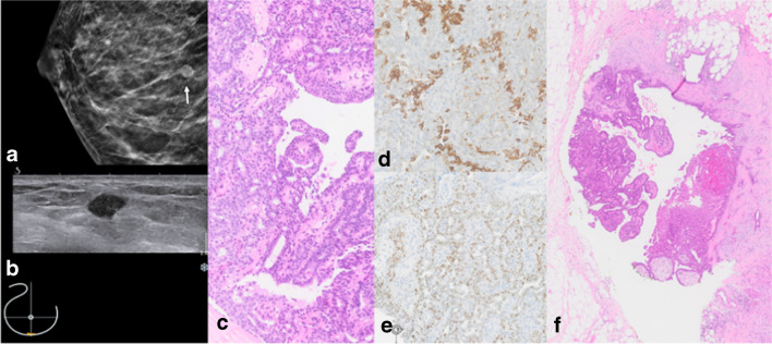
Fig. 4.
Papilloma without atypia. a Right cranio-caudal (cc) mammography image with hyperdense circumscribed mass lesion (white arrow). b Correlating small hypoechoic mass on ultrasound. c H&E stain of the core needle specimen shows papilla with a fibrous stroma and a heterogeneous mixture of cytologically bland ductal epithelium and myoepithelium. d CK5/6 mosaic pattern and e heterogeneous mostly weak to moderate ER expression. f Open excision (OE) specimen confirms benign papilloma

