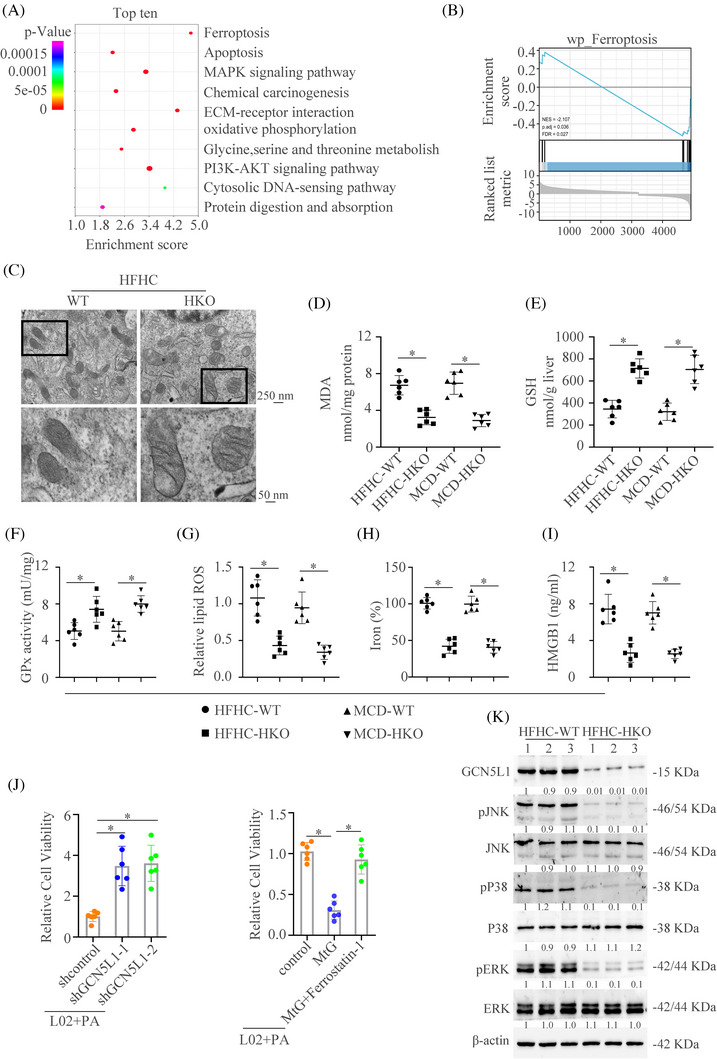
FIGURE 4.

GCN5L1 promotes ferroptosis of hepatocyte during NASH progression (A) KEGG analysis showed the top 10 pathway enrichment in GCN5L1 HKO group versus WT group. (B) Gene Set Enrichment Analysis (GSEA) showed the importance of ferroptosis in the GCN5L1 HKO group versus the WT group. (C) Transmission electron microscope showed the mitochondria in liver tissues. (D–F) Malondialdehyde (MDA), GSH, and GPx activity were assayed in WT and GCN5L1 HKO mice treated with HFHC or MCD. (G) Lipid ROS from isolated hepatocytes was measured by C11‐BODIPY staining coupled with flow cytometry. (H) Iron content was measured in WT and GCN5L1 HKO mice treated with HFHC or MCD. (I) HMGB1 was measured by ELISA in the serum of indicated groups. (J) Cell viability was assayed in different indicated cells. (K) Western blot was applied to detect the activation of the MAPK pathway.
