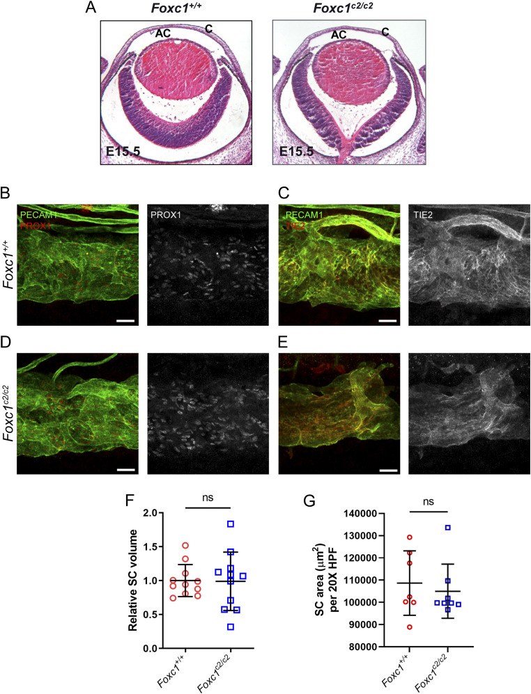
Figure 8. Substitution of Foxc2 into the Foxc1 locus does neither impair anterior eye segment development nor SC morphogenesis.
(A, E) Representative images of hematoxylin and eosin-stained transverse eye sections from embryonic day (E) 15.5 Foxc1+/+ and Foxc1c2/c2 mice show no difference in the normal development of the anterior chamber. AC, anterior chamber, C, cornea. (B, C, D, E) Representative images of CD31 and PROX1 (B, D) or Tie2 (C, E) expression in the SC of adult Foxc1+/+ (B, C) and Foxc1c2/c2 mice (D, E). Scale bars are 50 μm. (F) Relative SC volumes of Foxc1c2/c2 and Foxc1+/+ mice in a 1.5 mm × 1.5 mm field of view. SC volume for both groups was normalized by mean Foxc1+/+ SC volume. N = 11 volumes from 11 individuals for Foxc1+/+ and N = 11 volumes from 11 individuals for Foxc1c2/c2 mice. (G) Quantification of SC area per 20X high-power field. N = 7 for Foxc1+/+ and N = 8 for Foxc1c2/c2 mice. Data are mean ± SD. Statistical analysis: unpaired t test.
Source data are available for this figure.

