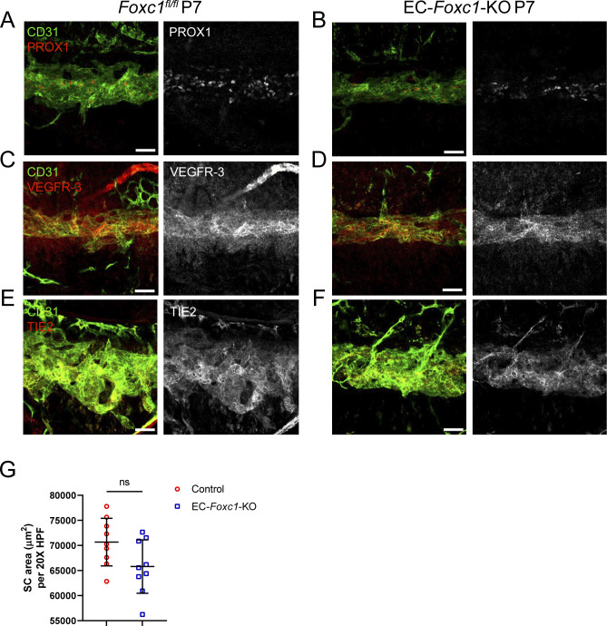
Figure S8. SC morphogenesis is not severely impacted by early postnatal, endothelial-specific deletion of Foxc1.
(A, B, C, D, E, F) Representative images of CD31 and PROX1 (A, B), VEGFR-3 (C, D) or Tie2 (E, F) expression in the SC of P7 Foxc1fl/fl control (A, C, E) and EC-Foxc1-KO mice (B, D, F). Scale bars are 50 μm. (G) Quantification of SC area per 20X high-power field in P7 Foxc1fl/fl control and EC-Foxc1-KO mice. N = 9 for Control and N = 9 for EC-Foxc1-KO mice. Data are mean ± SD. Statistical analysis: unpaired t test.
Source data are available for this figure.

