. 2023 May 4;13(7):1696–1719. doi: 10.1158/2159-8290.CD-22-1315

Figure 5.

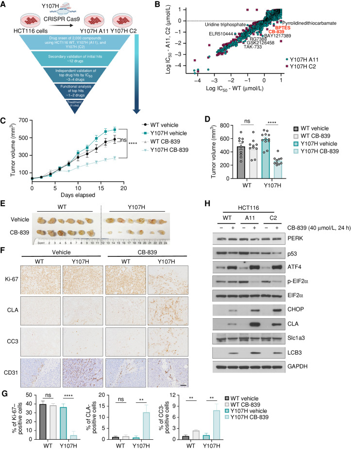
Figure 5. Y107H colorectal cancer cells show increased sensitivity to the glutaminase inhibitor CB-839. A, Schematic of CRISPR generation of HCT116 colorectal cancer cells with the Y107H mutation (clones A11 and C2) and subsequent screen for compounds that induce enhanced loss of viability in Y107H clones. (Created with BioRender.com.) B, Log IC50 of compounds against HCT116 cells with WT p53 or the Y107H clones A11 and C2. Compounds showing significantly increased sensitivity in both Y107H clones are indicated. C, Tumor growth of HCT116 cells with WT p53 or Y107H clone A11 in NSG mice (n = 10–12 per group) after treatment of vehicle or CB-839. After tumors reached 50 mm3, vehicle or CB-839 was administered 2× daily by oral gavage. Linear mixed model estimated difference in decreased tumor growth rate (mm3/day) by treatment with CB-839. ****, P < 0.0001; ns, not significant. D, Final tumor volume, shown as mean volume ± SEM of HCT116 tumors treated with vehicle or CB-839. ****, P < 0.0001; ns, not significant. E, Representative images of HCT116 tumors treated with vehicle or CB-839. F, Representative IHC from HCT116 tumors (n = 3–5 mice per condition) treated with vehicle or CB-839 and stained for the indicated antibodies. Scale bar, 50 μm. G, Percentages of positive cells of IHC HCT116 tumors. Averages ± SEM from at least three random images from n = 3–5 mice per condition. H, HCT116 cells were treated with 40 μmol/L CB-839 for 24 hours and assessed by Western blot for the indicated antibodies. **, P < 0.01; ****, P < 0.0001; ns, not significant, two-tailed unpaired t test with Welch correction.

Y107H colorectal cancer cells show increased sensitivity to the glutaminase inhibitor CB-839. A, Schematic of CRISPR generation of HCT116 colorectal cancer cells with the Y107H mutation (clones A11 and C2) and subsequent screen for compounds that induce enhanced loss of viability in Y107H clones. (Created with BioRender.com.) B, Log IC50 of compounds against HCT116 cells with WT p53 or the Y107H clones A11 and C2. Compounds showing significantly increased sensitivity in both Y107H clones are indicated. C, Tumor growth of HCT116 cells with WT p53 or Y107H clone A11 in NSG mice (n = 10–12 per group) after treatment of vehicle or CB-839. After tumors reached 50 mm3, vehicle or CB-839 was administered 2× daily by oral gavage. Linear mixed model estimated difference in decreased tumor growth rate (mm3/day) by treatment with CB-839. ****, P < 0.0001; ns, not significant. D, Final tumor volume, shown as mean volume ± SEM of HCT116 tumors treated with vehicle or CB-839. ****, P < 0.0001; ns, not significant. E, Representative images of HCT116 tumors treated with vehicle or CB-839. F, Representative IHC from HCT116 tumors (n = 3–5 mice per condition) treated with vehicle or CB-839 and stained for the indicated antibodies. Scale bar, 50 μm. G, Percentages of positive cells of IHC HCT116 tumors. Averages ± SEM from at least three random images from n = 3–5 mice per condition. H, HCT116 cells were treated with 40 μmol/L CB-839 for 24 hours and assessed by Western blot for the indicated antibodies. **, P < 0.01; ****, P < 0.0001; ns, not significant, two-tailed unpaired t test with Welch correction.