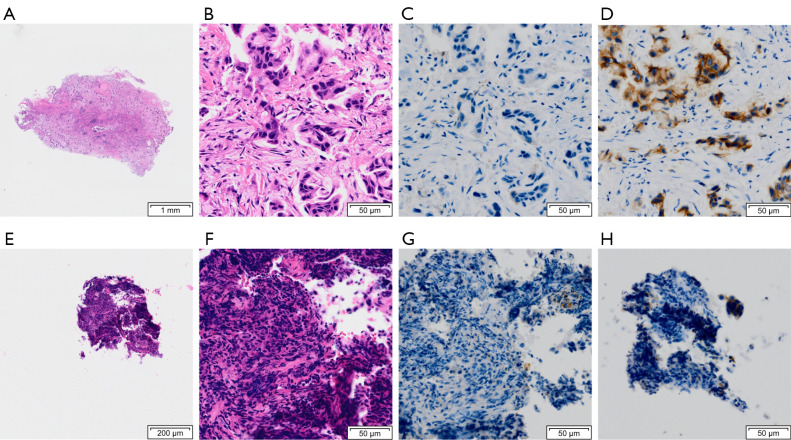
Figure 4.
A representative case of adenocarcinoma showing discordance in the results of the IHC scores for anti-HER3. The upper images show serial sections of cryobiopsy specimens (A ×1 magnification, B-D ×20 magnification) whereas the lower ones show those of forceps specimens (E ×2 magnification, F-H ×20 magnification). The sizes of the cryobiopsy and forceps biopsy specimens are 3.7 mm × 5.4 mm (A) and 0.8 mm × 0.6 mm (E), respectively. The cryobiopsy specimen retains a well-defined architecture (B) whereas the forceps biopsy specimen is somewhat crushed (F). The tumor proportion scores for programmed death-ligand 1 are both <1% (C,G). Although the IHC score for HER3 can be determined as 2+ for the cryobiopsy specimen (D), it is difficult to evaluate the IHC score for the forceps biopsy specimen due to loss of tumor cells during thin sectioning and is determined as 0 (H). IHC, immunohistochemistry; HER, human epidermal growth factor receptor.

