
FIGURE 6:
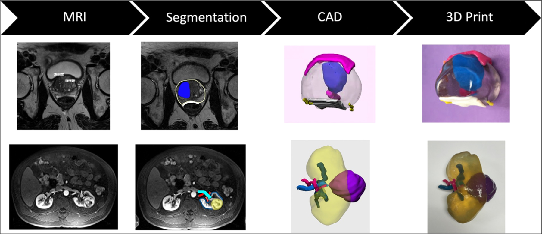
(Top Row) A 68-year-old male with a Prostate Imaging Reporting and Data System (PI-RADS) 5 lesion broadly abutting capsule without gross extraprostatic extension. In this case, the 3D printed model which included the prostate (clear), lesion (blue), bladder neck and urethra (pink), rectal wall (white), and neurovascular bundles (yellow) helped to facilitate a nerve sparing prostatectomy. (Bottom Row) A patient with a interpolar renal mass which abuts a lower pole calyx. The 3D printed model which included the kidney (transparent), mass (purple), artery (pink), vein (light blue), and collecting system (dark blue) allowed the vasculature to be well-visualized and facilitated a nephron sparing partial nephrectomy. For both cases, image segmentation and CAD modeling were performed in Mimics and 3-matic (Materialise, Leuven, Belgium) and 3D printing was performed using material jetting technology (J750 and Connex500, Stratasys, Eden Prairie, MN).