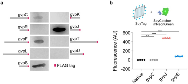
Figure 6. Genetic functionalization of bicones.
a) Representative images of dot blots. A FLAG tag (shown in red) was appended to the end of each GV gene and incorporation of the fusion protein into purified bicones was assessed using an anti-FLAG antibody. b) Top: Purified bicones, unmodified or with SpyTag appended to GvpC, gvpJ, or gvpS, were reacted with the fluorescent protein, SpyCatcher-mNeonGreen. Bottom: Mean fluorescence intensity of purified particles after conjugation. N = 4. Error bars, ± SEM. Welch’s t-test, (****, p < 0.0001; n.s, p ≥ 0.05).

