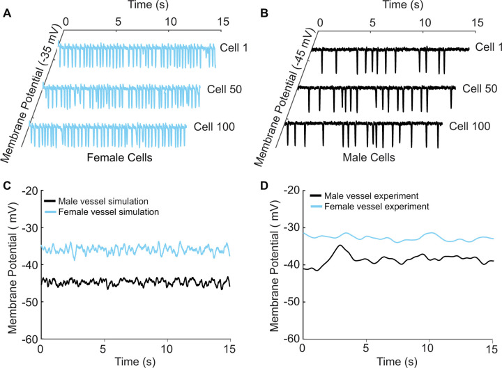
Figure 8. A one-dimensional tissue model representation of vascular smooth muscle cells connected in series.
A) Uncoupled female vessel simulation showing cell 1, cell 50, and cell 100 at a baseline membrane potential of −35 mV. B) Uncoupled male vessel simulations showing cell 1, cell 50, and cell 100 at a baseline membrane potential of −45 mV. C) Composite female (blue trace) and male (black trace) membrane potential of 400 coupled smooth muscle cells connected with gap junctional resistance of 71.4 Ωcm2 in a one-dimensional tissue representation. (D) Sharp-electrode records of the membrane potential of smooth muscle in pressurized (80-mmHg) female and male arteries from O'Dwyer et al., 2020.

