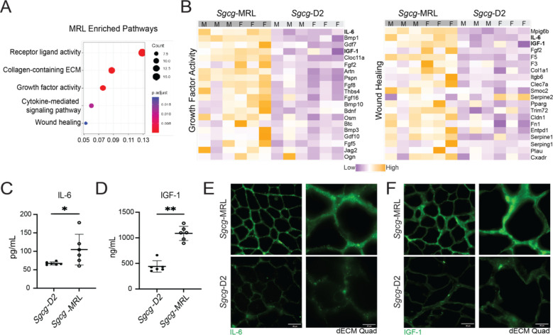
Figure 6. IGF-1 and IL-6 were upregulated in Sgcg-MRL serum.
Serum was collected from Sgcg-MRL and Sgcg-D2 cohorts at 20 weeks of age and evaluate using the SOMAscan aptamer assay. (A) Pathway enrichment analysis showed receptor ligand, collagen containing ECM and growth factor activity highly enriched in Sgcg-MRL serum. (B) Heatmaps of proteins in the growth factor activity (left) and wound healing (right) pathways upregulated in Sgcg-MRL mice. IL-6 and IGF-1 (bold) were among the most differentially expressed proteins in both pathways. IL-6 (C) and IGF-1 (D) upregulation was verified using ELISA analysis. (E) and (F) show matrix deposition of IL-6 and IGF-1 increased in decellularized myoscaffolds from Sgcg-MRL but not Sgcg-D2 muscles. Graphical quantification of mean ± SD. Statistical significance was determined using the Kolmogorov-Smirnov test. * p<0.05, ** p<0.01

