-
A, B
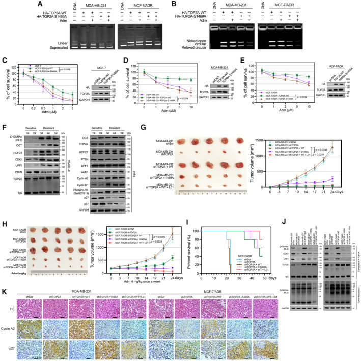
O‐GlcNAcylation attenuated Adm‐induced inhibition of TOP2A catalytic activity in DNA cleavage (A) and kDNA decatenation assays (B). TOP2A‐WT or TOP2A‐S1469A (20 nM) was immunoprecipitated using anti‐HA magnetic beads from breast cancer cells. 200 μM Adm was added during the reaction (30 min).
-
C–E
TOP2A‐WT or TOP2A‐S1469A overexpressed breast cancer cells were incubated with the indicated doses of Adm for 48 h. Cell viability was assessed with a CCK‐8 assay. Protein expression was analyzed by Western blot. n = 3 biological replicates. Paired t‐test was used for statistical comparison. P‐value was indicted. The data are presented as means ± SD.
-
F
TOP2A co‐IP was performed in five breast cancer patient tumor samples (chemotherapy‐sensitive, no. 1‐2; chemotherapy‐resistant or relapsed, no. 3‐5), and the immunoprecipitated fractions were analyzed by Western blot for the indicated proteins.
-
G
The effects of TOP2A O‐GlcNAcylation on tumor xenografts in nude mice. MDA‐MB‐231 cells with stable TOP2A silencing by shRNA (shTOP2A) were transfected with TOP2A‐WT or TOP2A‐S1469A expression plasmids. Cells were injected subcutaneously into the axillae of nude mice (n = 5 biological replicates for each group). 1 mg/kg L01 was administrated by tail vein injection. Volumes of tumors were monitored with caliper twice a week until 24 days. Paired t‐test was used for statistical comparison. P‐value was indicted. The data are presented as means ± SD.
-
H
In vivo antitumor performance of Adm in MCF‐7/ADR bearing nude mice. MCF‐7/ADR cells with stable TOP2A silencing by shRNA (shTOP2A) were transfected with TOP2A‐WT or TOP2A‐S1469A expression plasmids. Cells were injected subcutaneously into the axillae of nude mice (n = 5 biological replicates for each group). 4 mg/kg Adm and 1 mg/kg L01 was administrated. Volumes of tumors were monitored with caliper twice a week until 24 days. Paired t‐test was used for statistical comparison. P‐value was indicted. The data are presented as means ± SD.
-
I
Survival rates of MCF‐7/ADR bearing mice in different treatment groups within 48 d (n = 5 biological replicates).
-
J
TOP2A O‐GlcNAcylation and the interaction between TOP2A and cell cycle regulators were measured by co‐IP in tumor tissues. Cellular O‐GlcNAcylation was analyzed by Western blot.
-
K
IHC staining of Cyclin A2 and p27 in paraffin sections of MDA‐MB‐231 and MCF‐7/ADR tumors. The micrograph scale bar represents 50 μm.

