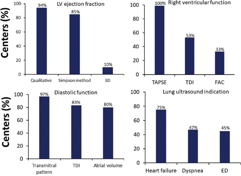
Figure 2.

Percentage of centers that evaluated: LV EF (top on the left) using qualitative method, Simpson method or with TTE – 3D; right ventricular function (top on the right) using TAPSE, TDI, and FAC, in 75 centers (33%); diastolic function (bottom on the left) using transmitral pattern, Doppler tissue imaging of mitral annulus (TDI) and left atrial volume, and principal indication of LUS (bottom on the right) in heart failure, dyspnea, and in hemodynamic instability in ED. LV EF = Left ventricular ejection fraction, TTE 3D = Transthoracic echocardiography three-dimensional, TAPSE = Tricuspid annular plane systolic excursion, TDI = Tissue Doppler imaging, FAC = Fractional area change, LUS = Lung ultrasound, ED = Emergency department
