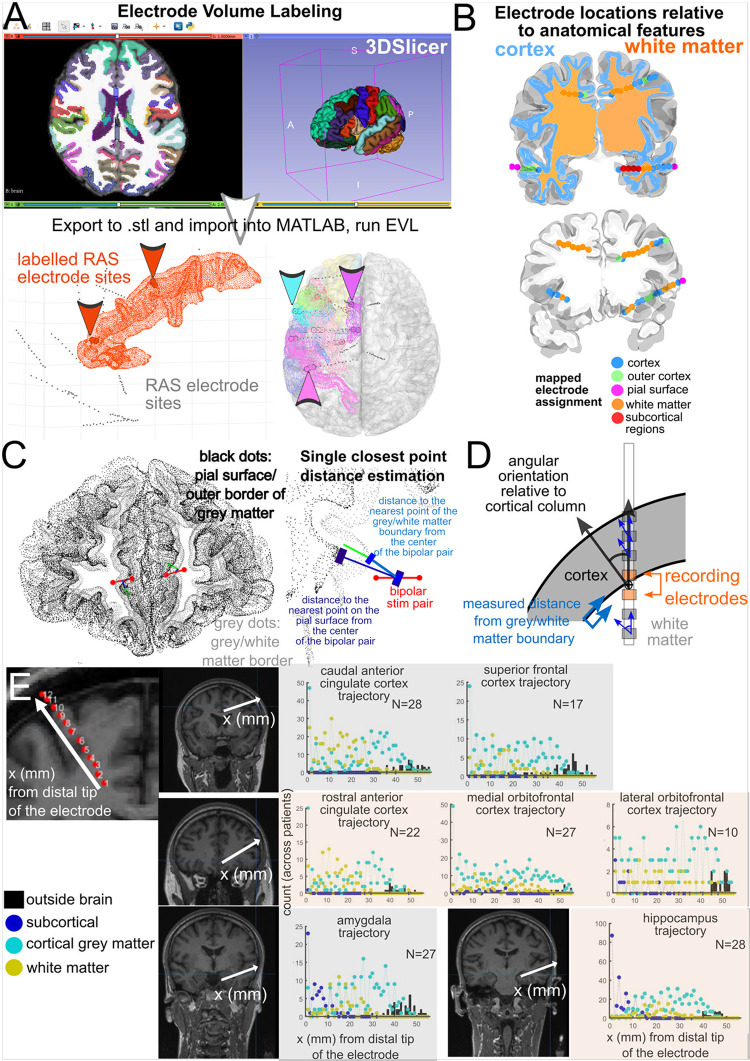
Fig 4. Electrode Volume Labelling and electrode location measurements.
A. Electrode Volume Labelling (EVL) steps, which involve exporting the aparc+aseg volume brain region labelling from FreeSurfer (such as from the DKT 40 map) to.stl files, which can be imported into MATLAB. Next, electrode contacts are labelled per brain region if they are contained within these volumes, which can contain the thickness of the cortical volume. B. Volumetric categorization of electrode contacts as white matter, cortex, subcortical structures, and outside the brain. C. Euclidean measurements of distances between the recording contacts and the nearest anatomical features. D. Further measurements of electrode contacts relative to different features of the nearest cortical structure. E. Categorization of electrode locations relative to neuroanatomical structures (cortical grey matter, subcortical structures, white matter, and outside brain) for depth electrodes with different ending trajectories for different brain regions. Counts of electrode locations relative to different classifications are across individuals (up to 27 patients).

