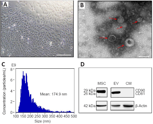
Figure 1.

Cell culture of MSCs from rat umbilical cord and collection of EVs.
(A) Eight days after cell culture of the Wharton’s Jelly from rat umbilical cords. The fusiform and polygon-shaped cells (MSCs) reached confluency in 8–10 days. Scale bar: 250 μm. (B) MSC-EVs under a transmission electron microscope. The red arrows indicate MSC-EVs. Scale bar: 500 nm. (C) Nanoparticle tracking analysis of MSC-EVs. (D) Protein expression of CD90 and CD81 (western blotting). CM: Culture medium (deprived of EVs); EV: extracellular vesicle; MSC: mesenchymal stem cell.
