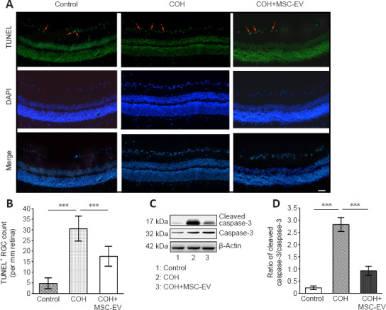
Figure 4.

Inhibition of apoptosis by MSC-EV treatment in the COH rat retina.
(A, B) TUNEL staining of the retina after COH and treatment with MSC-EVs. The TUNEL+ cells were stained green (red arrows, A). A significant increase in the number of apoptotic cells was observed in the ganglion cell layer in COH rats, while MSC-EV injection significantly prevented RGC apoptosis. Scale bar: 25 μm. (B) Quantitative results of TUNEL+ cells. (C) Caspase-3 and cleaved caspase-3 expression in the retina. (D) Quantitative analysis of the cleaved-caspase 3 expression in the retina. Data are expressed as the mean ± SD (n = 3/group). ***P < 0.001 (one-way analysis of variance followed by Tukey’s post hoc tests). COH: Chronic ocular hypertension; EV: extracellular vesicle; MSC: mesenchymal stem cell; TUNEL: terminal deoxynucleotidyl transferase-mediated dUTP nick-end labeling.
