Figure 2.
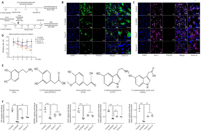
HucMSC-Exos co-localized with DA neurons and microglia in the lesioned substantia nigra improved behavior, and increased the concentrations of DA and its metabolites in lesioned striatum of PD model rats.
(A) Schematic diagram of the in vivo experiment. (B) Colocalization of Exos (red) and DA neurons (green), identified by TH expression, in the lesioned substantia nigra (n = 3). (C) Colocalization of Exos (red) and microglia (pink), identified by Iba-1 expression, in the lesioned substantia nigra. Blue indicates DAPI-stained nuclei (n = 3). The arrow indicates Exos, and the box indicates the enlarged area. Scale bars: 20 μm in B and C. (D) Intraperitoneal injection of APO (0.5 mg/kg) induced rotation in rats. Rotation was assessed 21 days after 6-OHDA injection (before Exos injection, i.e., 0 week) and 2, 4, 6, and 8 weeks after Exos injection. 6-OHDA group: injection of 6-OHDA without Exos; control group: no injection of 6-OHDA or Exos; Exos-LV group: 6-OHDA + Exos injected into the lateral ventricle; Exos-T group: 6-OHDA + Exos injected into the tail vein. **P < 0.01, vs. 6-OHDA group before injection (0 week), #P < 0.01, vs. before injection (0 week) (n = 6; repeated measures analysis of variance followed by Tukey’s post hoc test). (E) The chemical structure of all analytes including DA, DOPAC, HVA, 5-HT, and HIAA. (F) The levels of the metabolites were assayed by HPLC-MS. All data are shown as the mean ± SD. *P < 0.05, **P < 0.01, ***P < 0.001 (n = 3; one-way analysis of variance followed by least significant difference test). Exos-T and Exos-LV indicate 6-OHDA-induced PD models treated with Exos injection through the tail or lateral ventricle, respectively. 6-OHDA: 6-Hydroxydopamine; APO: apomorphine; DA: dopamine; DAPI: 4’,6-diamidino-2-phenylindole; Exos: exosomes; HPLC-MS: High performance liquid chromatography-mass spectrometry; hucMSCs-Exos: human umbilical cord mesenchymal stem cells derived exosomes; Iba-1: Ionized calcium binding adapter molecule 1; PD: Parkinson’s disease; TH: tyrosine hydroxylase.

