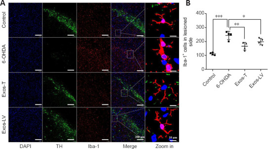
Figure 4.

HucMSC-Exos reduces microglial activation in the substantia nigra of PD model rats.
(A) Iba-1 (red) expression in the substantia nigra of rats from each group. Blue indicates DAPI-stained nuclei, and green (TH) indicates the substantia nigra region. Scale bars: 200 μm in the DAPI, TH, and Iba-1-stained images and the merge column and 10 μm in the last column. (B) Quantification of Iba-1 expression. The box indicates the enlarged area. All data are shown as the mean ± SD. *P < 0.05, **P < 0.01, ***P < 0.001 (n = 4, one-way analysis of variance followed by the least significant difference test). Exos-T and Exos-LV indicate the 6-OHDA-induced PD model treated with Exos injection through the tail or lateral ventricle, respectively. 6-OHDA: 6-Hydroxydopamine; DAPI: 4′,6-diamidino-2-phenylindole; Exos: exosomes; hucMSCs-Exos: human umbilical cord mesenchymal stem cells derived exosomes; Iba-1: ionized calcium binding adapter molecule-1; PD: Parkinson’s disease; TH: tyrosine hydroxylase.
