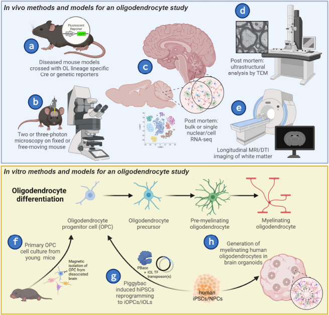
Figure 1.

Toolkit for studying oligodendrocytes (OLs).
(a–e) Tools for studying OLs in vivo. (a) Mouse models of disease can be crossed with OL lineage specific genetic reporters (i.e., fluorescent reporters) and Cre reporter lines for analysis of OL-specific perturbations. (b) Two/three-photon microscopy can be used in conjunction with OL-specific fluorescent reporter mouse models to explore longitudinal cellular activity and interactions. (c) RNA-sequencing (RNAseq) can be used to assess whole tissue or cell-specific transcriptional changes between control and disease brains or brain regions. (d) Myelination of axons can be quantified via transmission electron microscopy (TEM). (e) Changes in white matter can be observed overtime using magnetic resonance imaging (MRI) or diffusor tensor imaging (DTI). (f–h) Tools for studying OLs throughout maturation in vitro. (f) Isolating and culturing primary OPCs from young mice can be used to assess cell-autonomous dysfunction of OLs in disease at specific maturation states. Reprogramming human induced pluripotent stem cells (iPSCs) or neural progenitor cells (NPCs) into (g) induced OPCs (iOPCs) or induced oligodendrocytes (iOLs) or (h) generating myelinating OLs in brain organoids can be utilized to test OL dysfunction observed in animal models in a more human-relevant context. Created with BioRender.com.
