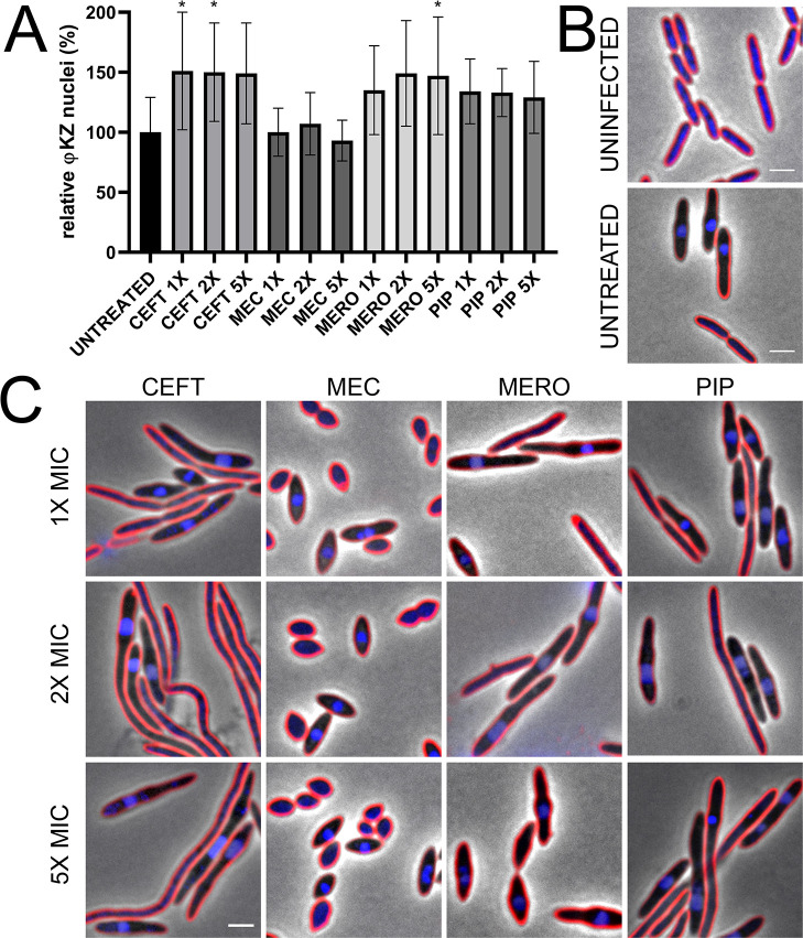
Fig 2. Treatment of P. aeruginosa K2733 with cell wall active antibiotics show differential φKZ infection related to MOA and host cell phenotype at 30 min post-infection.
(A) Quantification of phage infection (presence of distinct phage nuclei) under treatment conditions, relative to the untreated infected control. Error bars represent standard deviation of biological triplicates. * = p < 0.05 (B) Microscopy of uninfected and untreated infected controls, and (C) treated infected samples: ceftazidime (CEFT), mecillinam (MEC), meropenem (MERO), and piperacillin (PIP). Cell membrane strained with FM4-64 (red) and DNA stained with DAPI (blue). Scale bar represents 2 μm.

