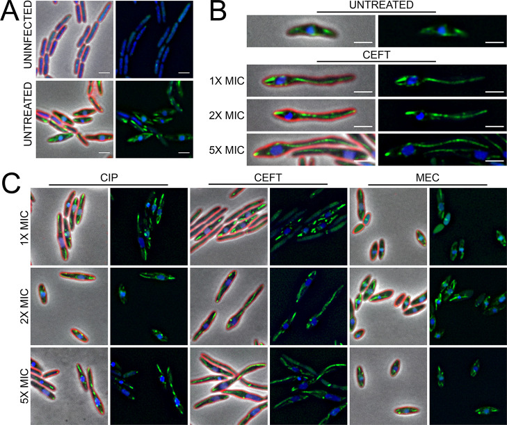
Fig 5. Antibiotic treatment leads to aberrant PhuZ filament and spindle dynamics at 30 min post-infection.
(A) Microscopy of uninfected and untreated infected controls. (B) Representative cell images of untreated infected controls, and CEFT treated infected samples at all concentrations. (C) Microscopy of treated infected samples. Cell membrane strained with FM4-64 (red) and DNA stained with DAPI (blue). GFP-PhuZ (green) under 0.1% arabinose induction. Scale bar represents 2 μm.

