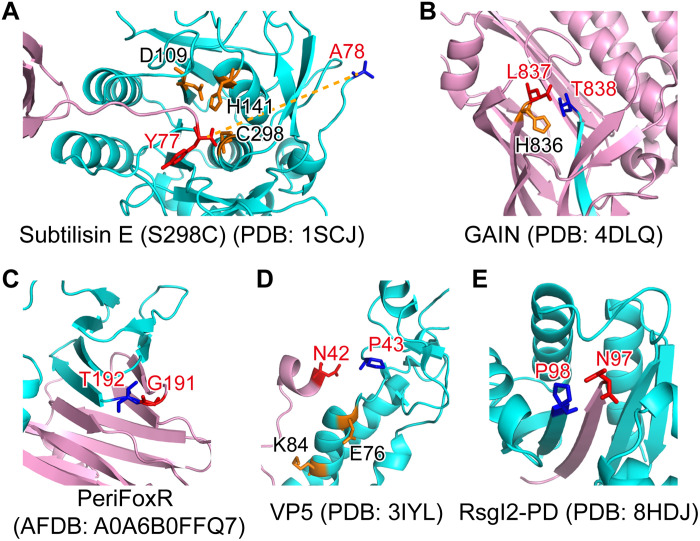
Fig. 3. The structure of RsgI-PD is distinct from other known autocleaved proteins.
The N- and C-terminal parts are colored pink and cyan, respectively. The P − 1 and P + 1 residues of the cleavage site are shown in red and blue sticks, respectively, and other residues probably involved in the catalysis are shown in orange sticks. (A) The structure of the propeptide-subtilisin E (S298C mutant) complex from B. subtilis. (B) The crystal structure of the GAIN domain of the GPCR CL1. (C) The AlphaFold2 structural model of the PD of FoxR (PeriFoxR) from P. aeruginosa. (D) The structure of the VP5 protein of Aquareovirus. (E) The structure of RsgI2-PD from C. thermocellum.

