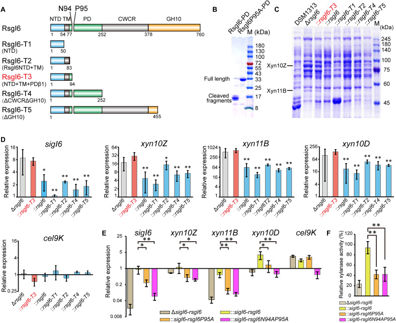
Fig. 4. Analysis of SigI6 and xylanase expression in mutant strains to demonstrate the role of autoproteolysis in signaling.
(A) Schematic representation of the composition of RsgI6 and its truncation mutants. RsgI6 contains an intracellular NTD, a TM, a PD, a proposed CWCR, and a C-terminal substrate-binding GH10 domain (GH10). (B) SDS-PAGE analysis of the purified SMT3-RsgI6-PD protein and its P95A mutant. (C) SDS-PAGE analysis of the extracellular proteins of wild type (DSM1313), ΔrsgI6, and its complemented strains with various RsgI6 truncations of C. thermocellum DSM1313, cultured in media with cellobiose as the carbon source. (D) qRT-PCR analysis of the genes of SigI6 and cellulosomal xylanases in ΔrsgI6 and its complemented strains grown in the cellobiose medium. The relative expression represents the ratio of transcribed mRNA levels in the mutant strains compared to that of DSM1313. The P values were calculated on the basis of three replicates using Student’s t test with ΔrsgI6::rsgI6-T3 as the reference. (E) qRT-PCR analysis of ΔsigI6-rsgI6 and its complemented strains grown in medium with alkali-pretreated wheat straw as carbon source. The relative expression represents the ratio of transcribed mRNA levels in the mutant strains compared to that of DSM1313. (F) Xylanase activity of the culture supernatant. The P values in (D) and (E) were calculated on the basis of three replicates using Student’s t test. *P < 0.05 and **P < 0.01.

