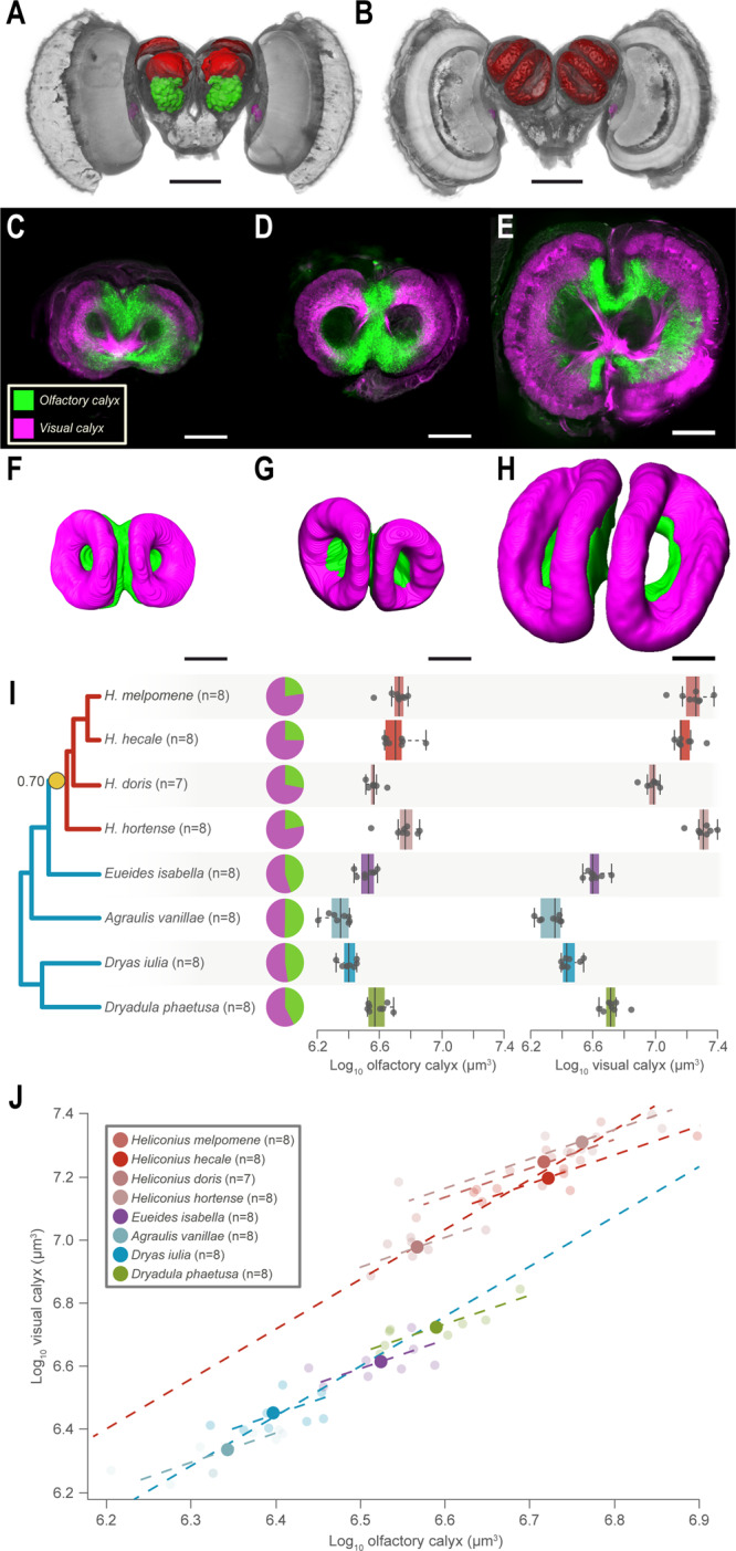
Fig. 4. Mushroom body expansion in Heliconius driven primarily by increased visual input.

A Anterior and B posterior 3D-reconstructions of H. hecale brain showing injection sites for the tracing of sensory projections to the MB (red neuropil). Olfactory projections neurons were traced from the antennal lobe (green neuropil) while visual projection were traced from injections around the ventral lobula (magenta). Scale bars in A and B = 500 μm. C Optical section of the mushroom bodies acquired with a confocal microscope in Dryas iulia, D Eueides isabella and E H. hecale. Inputs from olfactory (green) and visual (magenta) sensory neuropils terminate in segregated regions of the calyx. F–H 3D volumetric reconstructions of the visual and olfactory regions of the calyx in (F) Dryas iulia, (G) Eueides isabella and (H) H. hecale. Scale bars in C–H = 100 μm. I Heliconius exhibit a volumetric increase in the olfactory region of the mushroom body, but a greater increase in the visual region. The box encompasses two middle quartiles, with central line showing median. Whiskers extend to the furthest data point within 1.5 times the interquartile range. J Controlling for olfactory calyx volume, Heliconius exhibit an upshift in size of the visual calyx. Solid points = species means; faded points = individuals. Sample sizes are indicated in the figure ‘n’ = number of individuals/species. Source data files: I, J: Tracingdata.csv.
