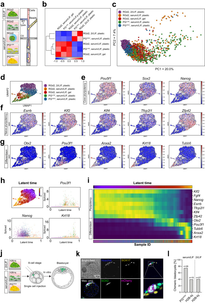
Fig. 6. Single-cell sequencing elucidates plakoglobin-induced re-establishment of naive pluripotency.
a Schematic of the different samples (RGd2 in serum/LIF in microgels and on plastic, PGlow and PGhigh cells in serum/LIF on plastic and naive control RGd2 cells in 2i/LIF on plastic) subjected to scRNA-seq analysis using the inDrop workflow. (RT: reverse transcriptase). b Pearson correlation heatmap of samples listed in (a). c Principal component (PC) analysis of samples listed in (a). d Uniform Manifold Approximation and Projection (UMAP) dimensional reduction plot of the scRNA-seq samples, for the maps of individual markers (in e–g). e Gene-expression values projected on the UMAP plot for core pluripotency markers (Pou5f1, Sox2, and Nanog). f Gene-expression values projected on the UMAP plot for naive pluripotency markers (Esrrb, Klf2, Klf4, Tfcp2l1, and Zfp42). g Gene-expression values projected on the UMAP plot for peri-implantation (Otx2 and Pou3f1) and serum-induced differentiation markers (Anxa2, Krt18, and Tubb6). h (top left) Latent time computation generated using scVelo and projected on the UMAP underlining the continuum between naive pluripotency (latent time = 0) and differentiation (latent time = 1). (top right) Single-cell spliced read counts arranged across the computed latent time for Pou3f1 (top right), Nanog (bottom left), and Krt18 (bottom right). i Heatmap representing the changes in gene expression across all cells arranged by latent time progression. j Schematic of single-cell injection into 8-cell stage embryos with subsequent in vitro culture until the blastocyst stage. k Confocal immunofluorescence image of a chimeric blastocyst that was injected at the 8-cell stage with a single serum/LIF cultured PGHIGH mESC. Blastocysts were stained for SOX2 (epiblast marker) and SOX17 (primitive endoderm marker). PGHIGH cells were identified by the PG-mCherry signal as shown in the merged image and highlighted by the white dotted line. Scale bar: 50 µm. l Chimeric blastocyst contribution efficiency of PGHIGH cells in serum/LIF (N = 26 embryos) and control H2B-tq cells in naive (2i/LIF; N = 27 embryos) and metastable pluripotent (serum/LIF; N = 15 embryos) conditions. Single cells were injected at the 8-cell stage followed by in vitro culture until blastocyst stage.

