Fig. 2.
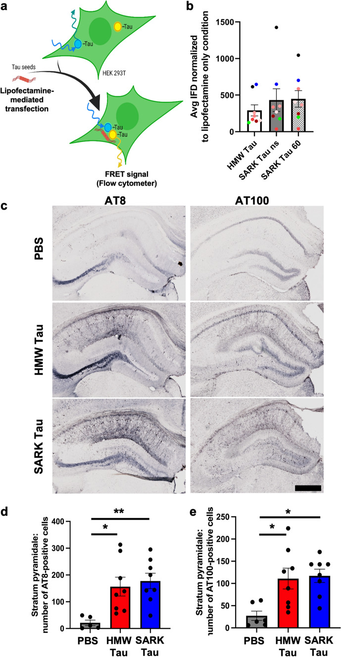
Tau oligomers and tau fibrils have similar bioactivities in vitro and in vivo. a Schematic of the FRET-biosensor seeding assay. HEK293T cells stably express the tau repeat domain fused to either CFP or YFP. When transfected with bioactive tau seeds, these constructs aggregate and become close enough to generate a FRET signal quantifiable by flow cytometry. Here, SARK and HMW tau samples from eight AD cases were normalized to total tau amounts (8 ng monomer equivalent per well) before being added to the cells. Artwork was created using BioRender. b Quantification of the seeding activity by flow cytometry 24 h after transfection. Integrated FRET densities were normalized to the lipofectamine only negative control. Data represented as mean ± SEM from three independent experiments, one color per case, Wilcoxon matched-pairs test, non-significant. Cases further used for in vivo experiments have been highlighted in green for #1892 and blue for #2399. ns non-sonicated; 60 = sonicated 60 pulses. c Representative images of phosphorylated AT8 and AT100 tau pathology in the dorsal hippocampus of injected PS19 mice 3 months after injection. Scale bar = 500 µm. d Quantification of the number of AT8-positive cells in the pyramidal layer (Stratum pyramidale) of the dorsal hippocampus of PS19 mice 3 months after injection. e Quantification of the number of AT100-positive cells in the pyramidal layer (Stratum pyramidale) of the dorsal hippocampus of PS19 mice 3 months after injection. Data represented as mean ± SEM, Kruskal–Wallis, Dunn’s multiple comparison, *p < 0.05

