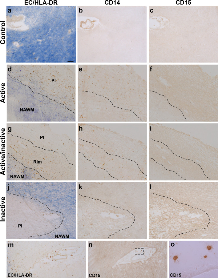
Fig. 1.
Expression pattern of CD14 and CD15 in MS lesions. a–c CD14 or CD15 staining was not detected in control human tissue. CD14+-cells were located both in the plaque of AL (d, e) and in the rim of AIL (g, h). By contrast, these cells were almost absent in the center or plaque of AIL (g, h) and IL (j, k). CD15 staining was not detected in any region of the MS lesions (f, i, l), although CD15+ granulocytes were clearly detected in the perivascular area of blood vessels (m–o). EC eriochrome cyanine, AL active lesions, AIL mixed active/inactive lesions, IL inactive lesions, Pl plaque, NAWM normal appearing white matter. N = 46 ALs, 26 AILs and 24 ILs from 33 MS patients Scale bar: a–c, j–n = 100 µm; d–i = 125 µm; o = 15 µm

