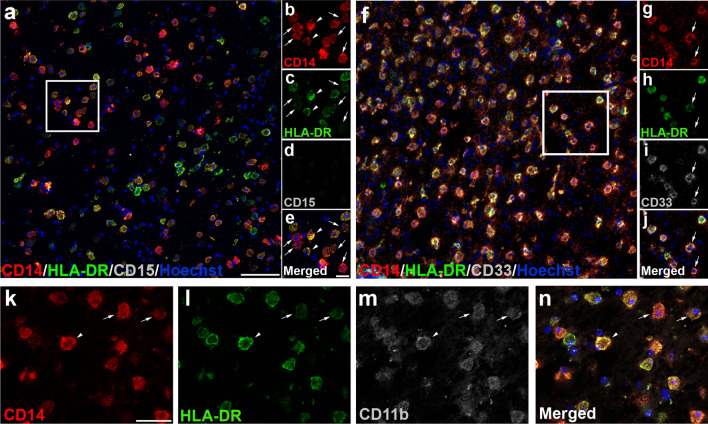
Fig. 2.
Immature myeloid monocytic cells are present within MS lesions. a Based on the intensity of HLA-DR, we identified different CD14+-cell subpopulations: CD14+HLA-DR−/low-cells were considered as M-MDSC-like cells (arrows in b–e) and CD14+HLA-DRhi-cells were identified as inflammatory macrophages (arrowheads in b–e). The lack of CD15 expression in CD14+-cells (d) corroborated the exclusive presence of M-MDSC-like cells. f–g CD33 as typical marker for immature myeloid cells was observed in CD14+HLA-DR−/low (arrows in g–j). k–n CD14+-cell subpopulations were identified as myeloid cells by CD11b staining. Arrows point to M-MDSC-like cells (CD14+HLA-DR−/low-cells). Arrowheads indicate CD11b+ inflammatory macrophages (CD14+HLA-DRhi-cells). N = 46 ALs, 26 AILs, and 24 ILs from 33 MS patients. Scale bar: a, f = 100 µm; insets in b–d = 20 µm, insets in g–j = 30 µm; k–n = 50 µm

