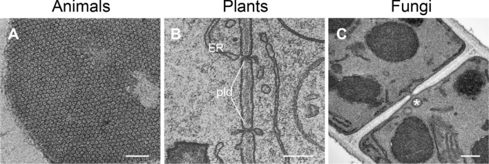
Fig. 3.
Transmission electron micrographs of cell–cell connections in animals, plants and fungi. A Cluster of an isolated junctional plaque from rat liver showing the gap junctions in a transversal cut. Adapted with permission from Ref. [84]. Scale bar 0.05 µm. B Plasmodesmata (pld) in a maize (Zea mays) root tip, with the endoplasmic reticulum (ER) spanning through the cell wall and linking neighboring cells. Adapted with permission from Ref. [124]. Scale bar, 0.1 µm. C Porous septum between two supracellular compartments of a Zymoseptoria tritici hypha. Note the Woronin body (*) (see Sect. “Organelles”) positioned right in front of the septal pore. Courtesy of Gero Steinberg, University of Exeter, U.K. Scale bar, 0.2 µm

