Fig. 4.
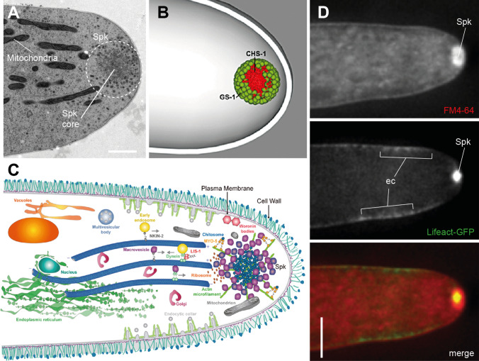
The fungal Spitzenkörper serves as an organizing center for hyphal tip growth. A A transmission electron micrograph section of the hyphal tip apex reveals the Spitzenkörper (Spk) as a dense vesicle cluster comprising a core with a surrounding vesicle cloud. Mitochondria are recruited into the apex to provide the huge amounts of energy required for fast tip growth. Image reproduced with permission from Ref. [169]. Scale bar, 500 nm. B The three-dimensional model of the functionally stratified Spk shows the glucane and chitin synthases GS-1 and CHS-1, respectively, as one of its main constituents (reproduced with permission from Ref. [214]). C Simplified schematic representation of the highly complex fungal tip growth machinery in which the Spk acts as a vesicle relay station: secretory vesicles deliver building blocks for plasma membrane and cell wall biosynthesis along microtubules towards the Spk. From there, vesicles are guided along F-actin tracks for targeted exocytosis to the apical plasma membrane to drive polarized hyphal tip extension. Not-incorporated material becomes reused through a subapical endocytic collar allowing for the very high tip extension rates seen in fungi (reproduced with permission from Ref. [165]). D Co-imaging of the fluorescent membrane marker FM4-64 with the F-actin reporter Lifeact-GFP shows the close functional relationship between exo- and endocytosis dynamics (ec endocytic collar) with the actin cytoskeleton (reproduced with permission from Ref. [14]). Scale bar, 5 µm. Please find further details on the topic in [62]

