
Fig. 7.
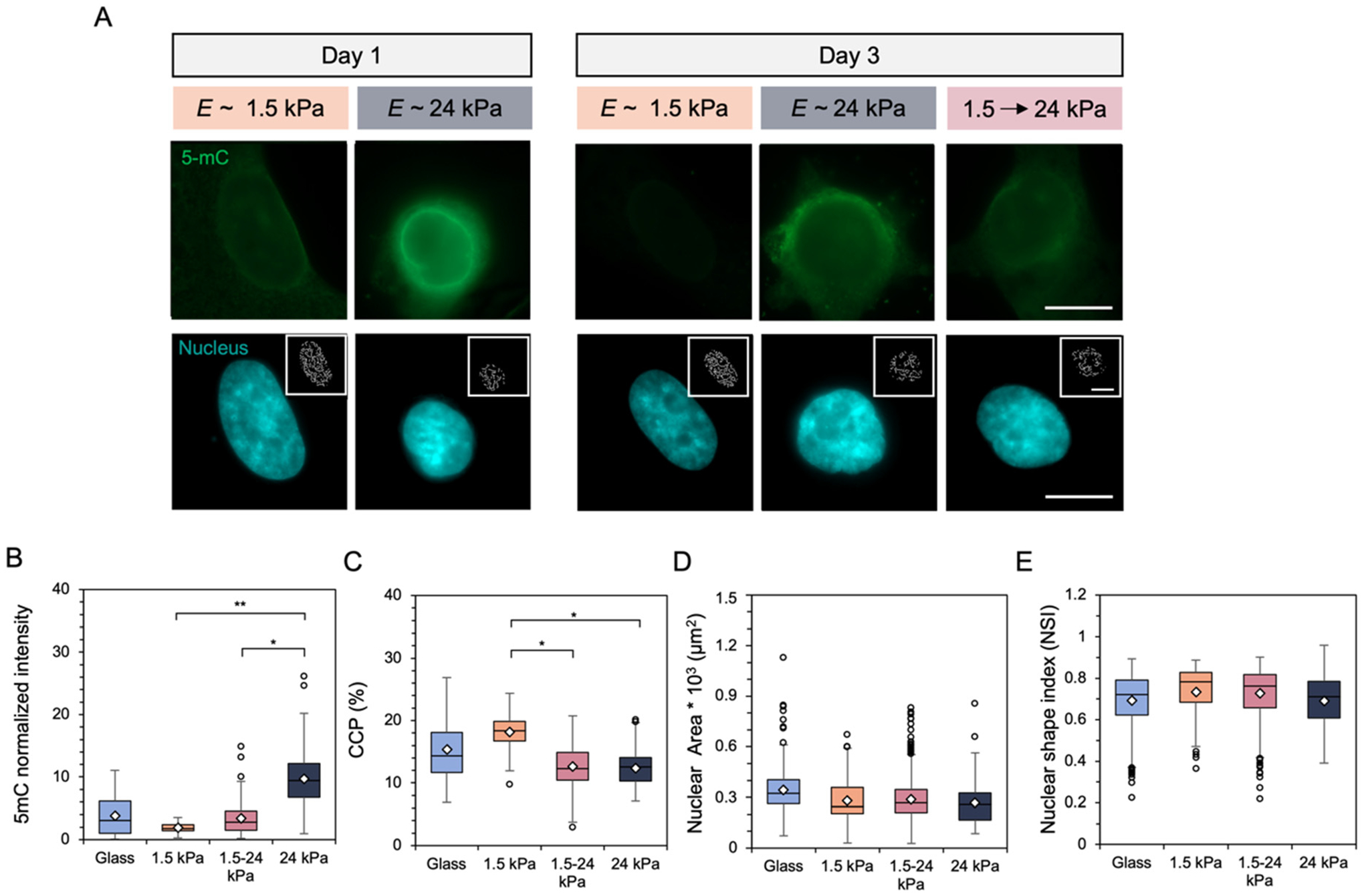
(A) Representative images of fibroblast global DNA methylation as indicated by 5-mC (green) staining and nuclei (DAPI) after (left) 1 day of culture on E ~ 1.5 or 24 kPa mechanically static hydrogels and (right) after 3 days on either the mechanically static hydrogels or a hydrogel that was stiffened from 1.5 to 24 kPa after 1 day. Inset images represent pixelated edges within the nuclei, used to quantify chromatin condensation percentage (CCP). Scale bars: 10 μm. Nuclear metrics were measured by (B) global DNA methylation intensity within the nucleus (C) the CCP, (D) nuclear spread area, and (E) nuclear shape index, which measures nuclear roundedness. N = 3 hydrogels per group. **P < 0.01, *P < 0.05.