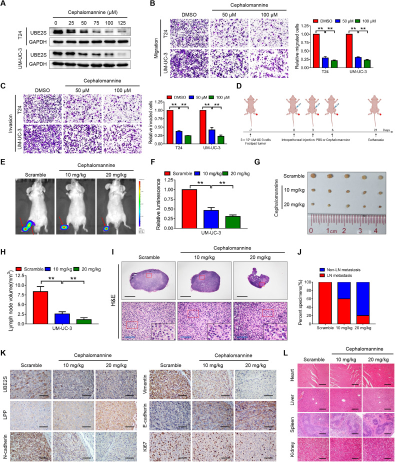
Fig. 7. Cephalomannine pharmacologically inhibits UBE2S and blocks the lymphatic metastasis of BCa cells.
A UBE2S protein levels in BCa cells treated with various concentrations of cephalomannine for 48 h, as determined by western blot assays. Representative images and quantitative analyses of transwell migration (B) and invasion (C) assays of BCa cells treated with cephalomannine at the indicated concentrations. D Schematic illustration of the intraperitoneal injection of PBS or cephalomannine in a UM-UC-3 popliteal lymphatic metastasis model. Representative bioluminescence images (E) and quantitative analysis (F) of popliteal metastatic LNs from nude mice. LNs lymph nodes. Representative images of popliteal LNs (G) and quantitative analysis of their volumes (H). I Representative images of H&E staining in popliteal LNs from each group. Scale bars: black, 500 μm; blue, 50 μm. J Percentages of lymphatic metastasis in each group. K Representative images of IHC staining showing UBE2S, LPP, N-cadherin, Vimentin, E-cadherin and Ki-67 expression in footpad tumors of the indicated groups. Scale bars: black, 50 μm. L Representative images of H&E staining in the major organs treated with the indicated concentrations of cephalomannine. Scale bars: black, 50 μm. **P < 0.01.

