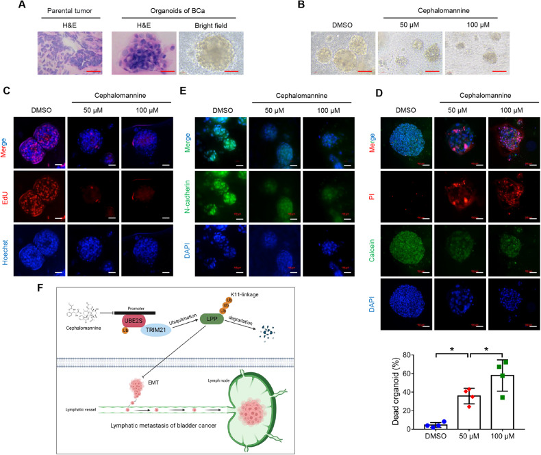
Fig. 8. Cephalomannine inhibits the growth and metastasis of organoids derived from human BCa.
A Representative images of H&E staining of parental tumors and PDOs and bright-field images of organoids. PDOs patient-derived organoids. Scale bars: red, 100 μm. B Bright-field images of BCa organoids treated with the indicated concentrations of cephalomannine for 96 h. Scale bars: red, 100 μm. C Measurement of the organoids in S phase using EdU assays. Scale bars: white, 100 μm. Hoechst was used to label the cell nucleus. D Representative images and quantitative analysis of cell viability in four BCa organoids treated with the indicated concentrations of cephalomannine for 96 h. PI was used to label the dead cells, while calcein was used to label the living cells. Scale bars: white, 100 μm. *P < 0.05 and **P < 0.01. E Representative immunofluorescent images showing N-cadherin expression in BCa organoids treated with the indicated concentrations of cephalomannine for 96 h. Scale bars: white, 100 μm. F Schematic diagram depicting the underlying mechanism of UBE2S function in lymphatic metastasis of BCa.

