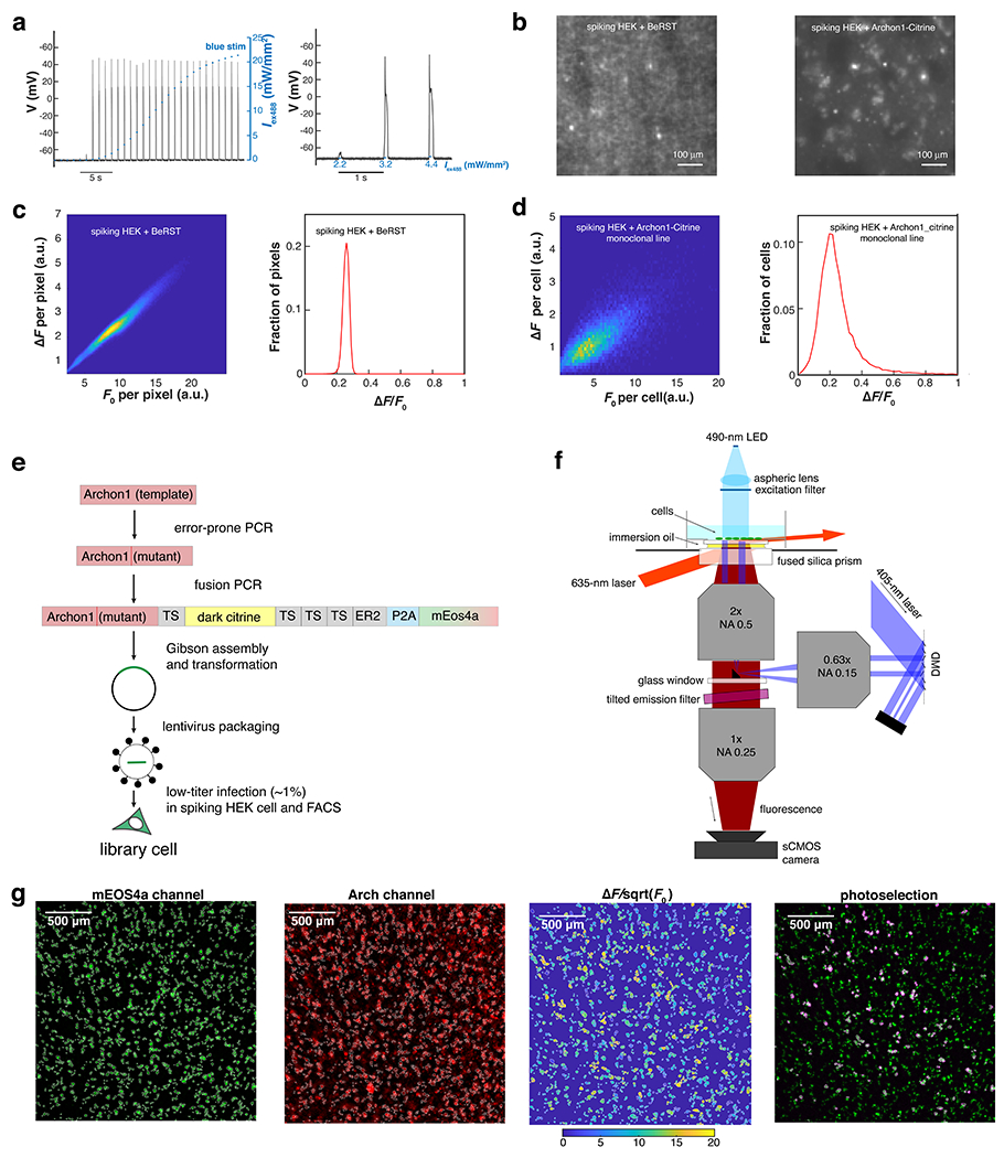
Extended Data Fig. 2. Video-based pooled screen for mutations that enhance the performance of Arch-derived GEVIs (Related to Fig. 2).

a. Current clamp measurement of membrane potential in spiking HEK cells reveals “all-or-none” spiking in response to increasing optogenetic stimulation (n = 2 trials; exc. 488 nm). Left: membrane potential in response to optical stimuli of increasing strength (0 - 22 mW/mm2). Right: enlarged view showing the threshold transition. b. Fluorescence image (exc. 635 nm) of spiking HEK cell monolayer stained with BeRST1 (left) or expressing Archon1-Citrine (right). In the Archon1-Citrine image, the presence of the spacer cells (spiking HEK cells that did not express Archon1-Citrine) enabled individual cells to be resolved. c. Distribution of membrane potential changes in a spiking HEK cell monolayer, reported via imaging of a voltage-sensitive dye BeRST1, plotted for each pixel. Left: heatmap of ΔF vs. F0 for all pixels in a 2.3×2.3 mm FOV (500×500 pixels). Right: histogram of ΔF/F0. The distribution had a fractional width (S.D./mean) of 8% (mean 0.25, S.D. 0.02; 99th percentile: 0.29). d. Distribution of Archon1 baseline brightness (F0) and voltage sensitivity (ΔF/F0) in a monoclonal Archon1-expressing spiking HEK cell monolayer, plotted for each cell (n = 20900 cells). Left: heatmap of ΔF vs. F0 for all cells in a 2.3 × 2.3 mm FOV (500×500-pixels). Right: histogram of ΔF/F0. The distribution had a fractional width (S.D./mean) of 43% (mean 0.23, S.D. 0.10; 99th percentile: 0.54), substantially broader than the distribution for BeRST1. e. Work-flow for the generation of the library cells. f. Optical system for video-based pooled screening. g. Image analysis for a representative FOV (the same as shown in Fig. 2e,f). The example was, from left to right: 1) ROIs generated by “Watershed” image segmentation in the mEos4a channel (exc: 490 nm; EGFP emission filter). 2) Baseline fluorescence (F0) image in the Arch channel (exc: 635 nm; Arch emission filter). 3) Heatmap of for individual ROIs. Here is used as a proxy for shot noise-limited for SNR. 4) Overlay of the patterned violet light (pseudo-color red; exc. 405 nm; CFP emission filter) and mEos4a image (exc: 490 nm; EGFP emission filter).
