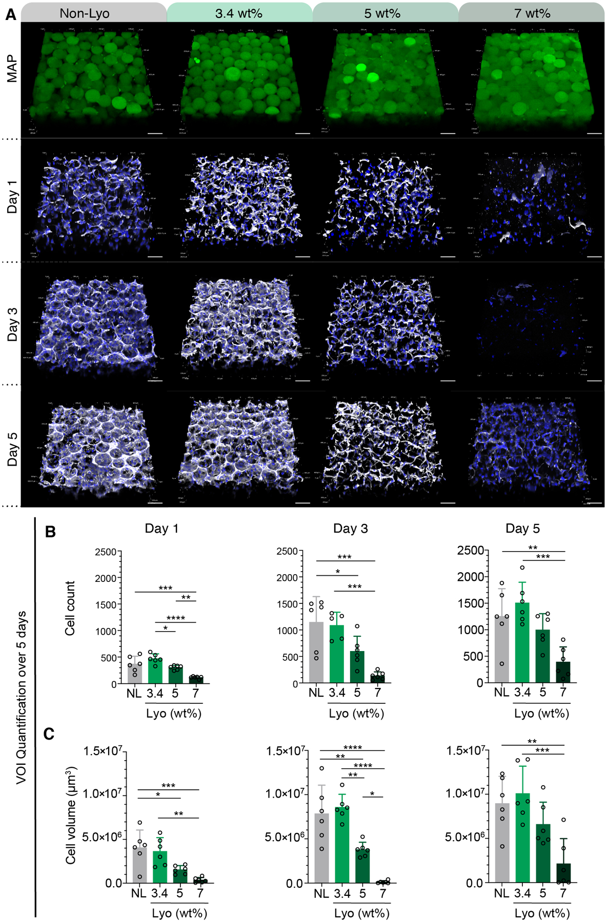
Figure 7.

(A) Split channels of confocal Z-stacks for MAP (green) composed of non-lyo-microgels and lyo-microgels reconstituted at 3.4, 5, and 7 wt% cultured with D1 cells (white F-actin, blue DAPI). Scale bar = 100 μm. Quantification of volume of interest (VOI) within each replicate across days 1, 3, 5 for (B) cell count and (C) cell volume as measured by volume renderings in IMARIS software of F-actin staining (white). N=3 with duplicate wells represented by individual datapoints. A one-way ANOVA with Tukey HSD was performed on the samples at each time point with significance reported at p < 0.05 (*), <0.01 (**), <0.005 (***), and <0.001 (****).
