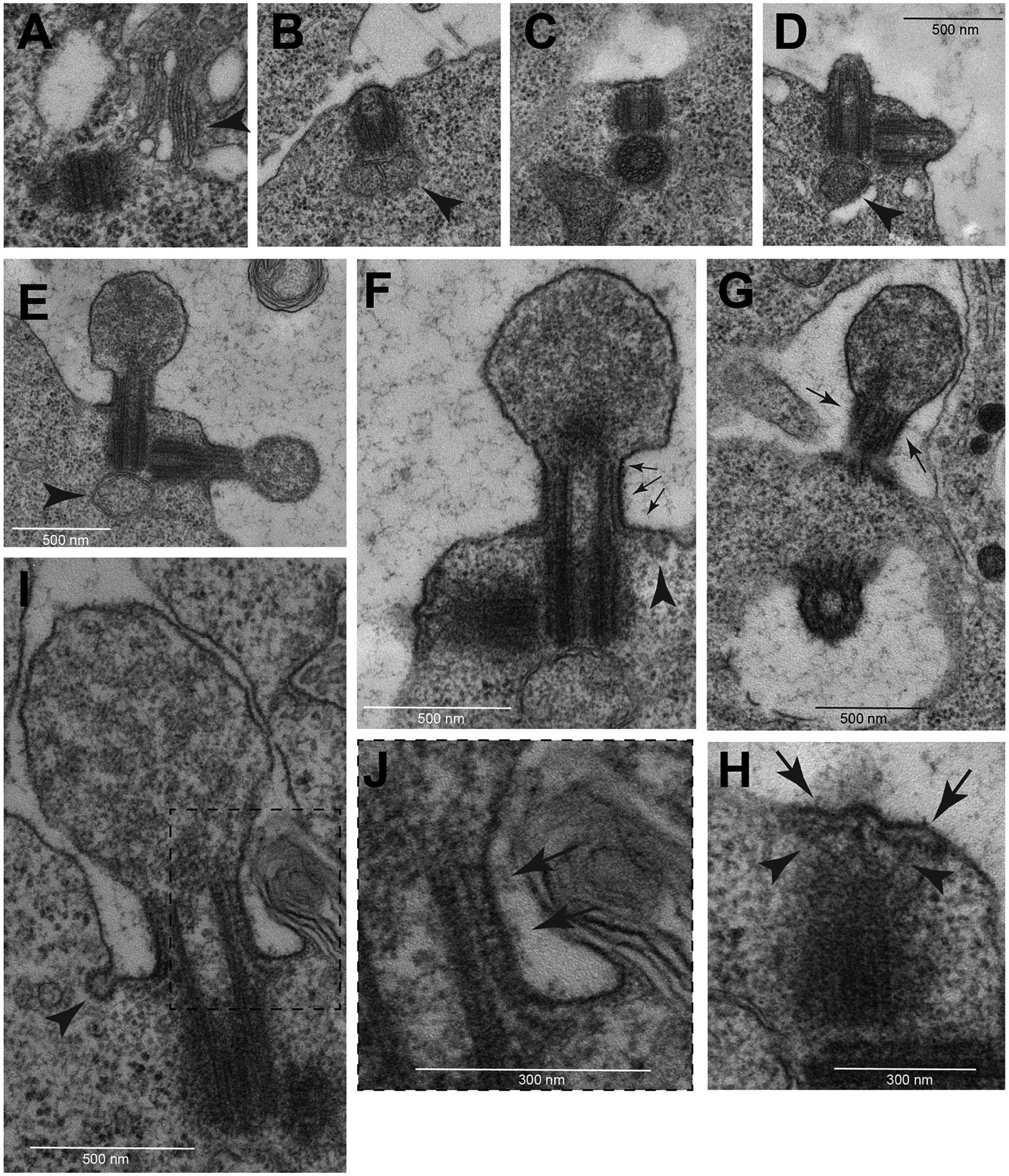
Fig. 2.

TEM images of early assembly stages of spermatocyte cilia. (A) At beginning of prophase the centrioles are located near the nucleus in association with Golgi cisternae (arrowhead). During leptotene the centrioles dock to the cell membrane (B) and form small buds (C) that elongate progressively during zygotene (D). During pachytene the distal region of the elongating cilia swell (E). One small mitochondrion is associated with the base of the mother centrioles (arrowheads in B,D,E). (F) L-shaped filamentous structures run from the base of the ciliary membrane toward the distal region of the cilium (arrows). (G) Grazing section of the CLR showing parallel bundles of these L-shaped filamentous structures (arrows). (H) Detail of the daughter centriole seen in (F) showing the sub membranous filaments (arrows) and distinct radial filaments emerging from the apical region of the centriole (arrowheads). (I) Early stage cilium and detail of its distal region (boxed region shown in J) showing regularly spaced links (arrows) connecting the ciliary membrane and the filamentous structures. Small vesicles are found at the base of the early cilia close to the cytoplasmic ends of the filamentous structures (arrowheads in F,I).
