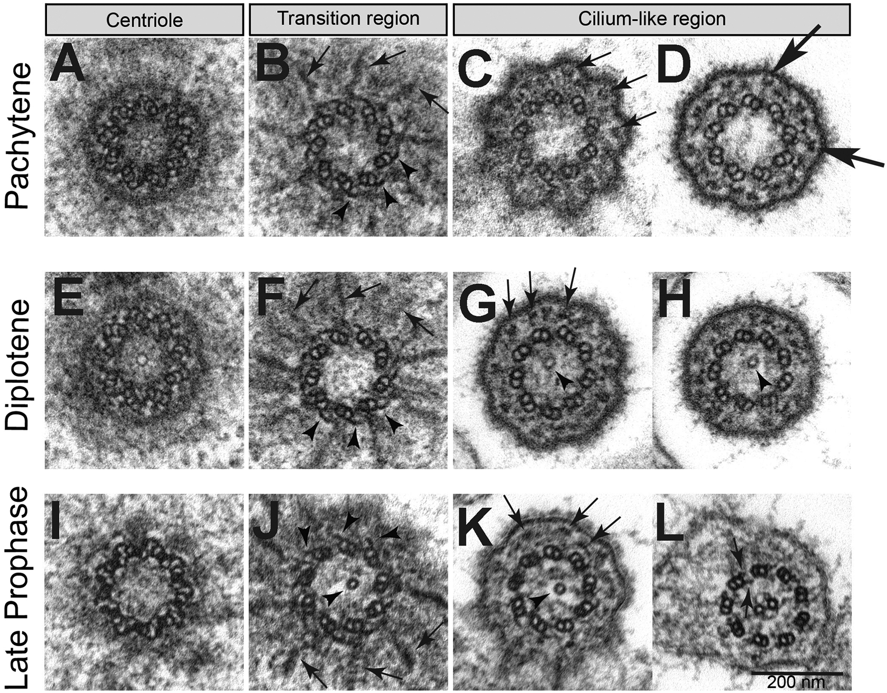
Fig. 3.

Features of centrioles and cilia during primary spermatocyte meiotic progression. Cross sections during (A-D) pachytene, (E-H) diplotene, (I-L) late prophase. (B,F,J) Radial filamentous structures (arrows) are seen at the transition zone between the distal region of the centrioles and the beginning of the ciliary axoneme; the C-tubules reduced to short hook-like structures (arrowheads). A single tubule is present in the lumen of the basal body and/or transition zone (arrowheads, G,H,J,K). (C,G,K) The filamentous structures running close to the ciliary membrane appear in cross sections as small aggregates of electron-dense material (arrows). (D) Y-linkers connect the A-tubule with the plasma membrane of the basal region of the cilium (large arrows). (L) Late primary spermatocytes display a ciliary axoneme consisting of a canonical 9+2 model with dynein arms (arrows).
